| |
|
| 
|
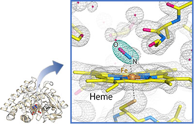
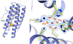
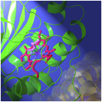
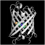
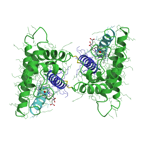
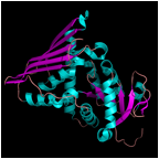
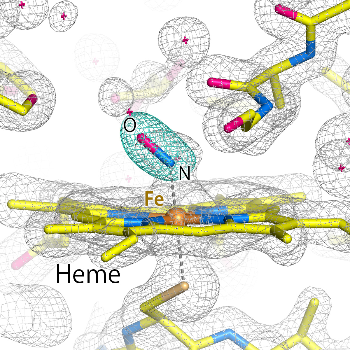
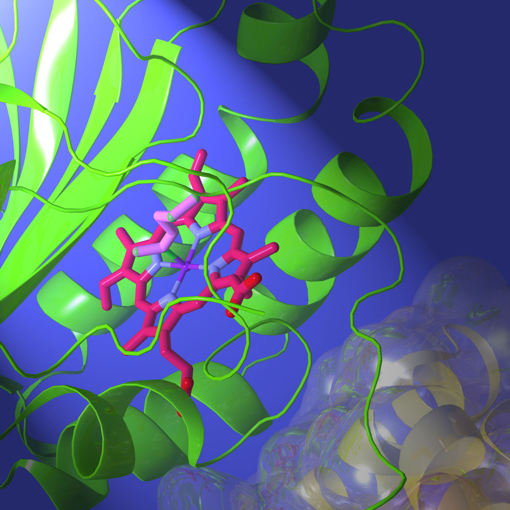
NO reductaseの反応中間体 (P450nor)
- "Short-lived intermediate in N2O generation by P450 NO reductase captured by time-resolved IR spectroscopy and XFEL crystallography", Proc. Nat. Acad. Sci. ,118, e2101481118 (2021)
- 温室効果・オゾン層破壊の原因である亜酸化窒素の生物的発生機構の解明- プレスリリース (2021年5月18日)
|
| 
|
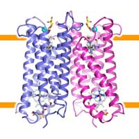
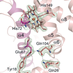
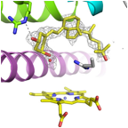
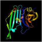
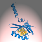
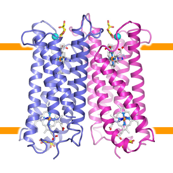
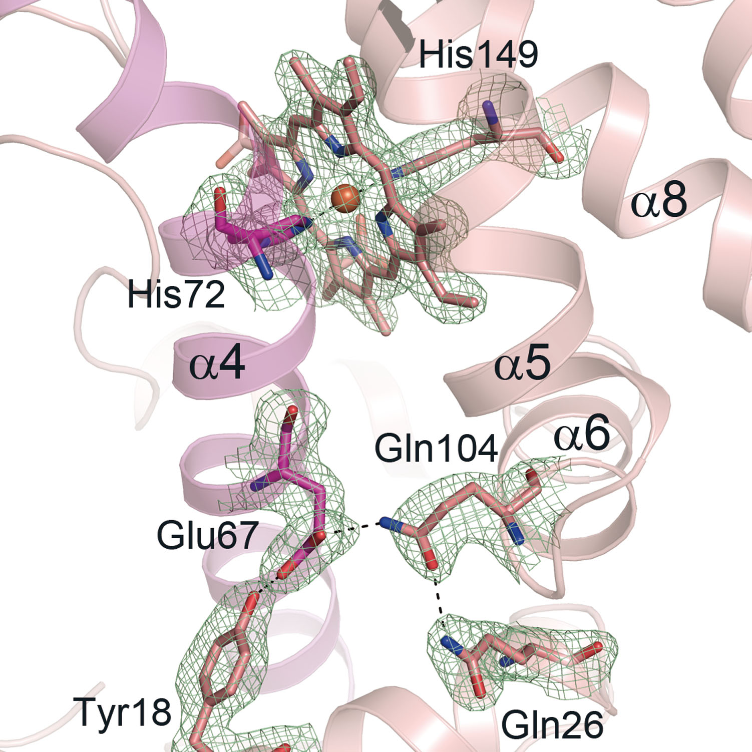
ヒト小腸で鉄分吸収に関わる膜タンパク質 (Dcytb)
- Ganasen, M., Togashi, H., Takeda, H., Asakura, H., Tosha, T., Yamashita, K., Hirata, K., Nariai, Y., Urano, T., Yuan, X., Hamza, I., Mauk, A. G., Shiro, Y., Sugimoto, H., Sawai, H. "Structural basis for promotion of duodenal iron absorption by enteric ferric reductase with ascorbate". Commun. Biol. 1, 120 (2018) [doi]
- 貧血予防の新たな指針へ向けて -ビタミンCが鉄分の吸収を促進するメカニズムを原子レベルで解明- プレスリリース (2018年8月20日)
|
| 
|
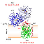
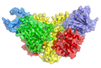
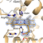
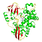
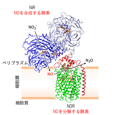
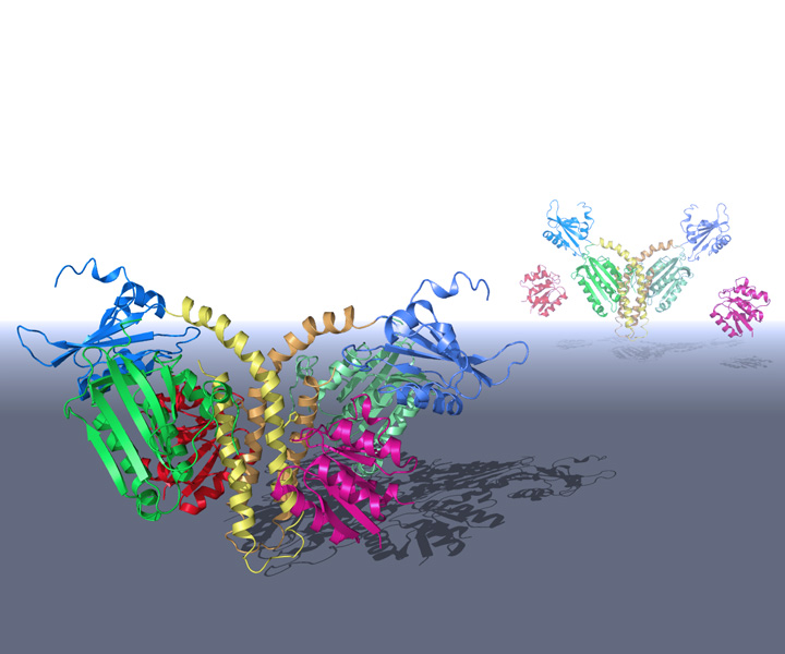
脱窒酵素の超分子複合体 (NiR-cNOR complex)
- Terasaka, E., Yamada, K., Wang, P. H., Hosokawa, K., Yamagiwa, R., Matsumoto, K., Ishii, S., Mori, T., Yagi, K., Sawai, H., Arai, H., Sugimoto, H., Sugita, Y., Shiro, Y., Tosha, T.: "Dynamics of nitric oxide controlled by protein complex in bacterial system" Proc. Natl. Acad. Sci. U.S.A. (2017) [doi]
- 合成酵素と分解酵素の協演- プレスリリース (2017年8月29日)
|
| 
|
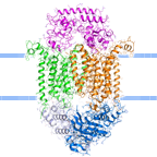
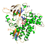
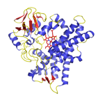
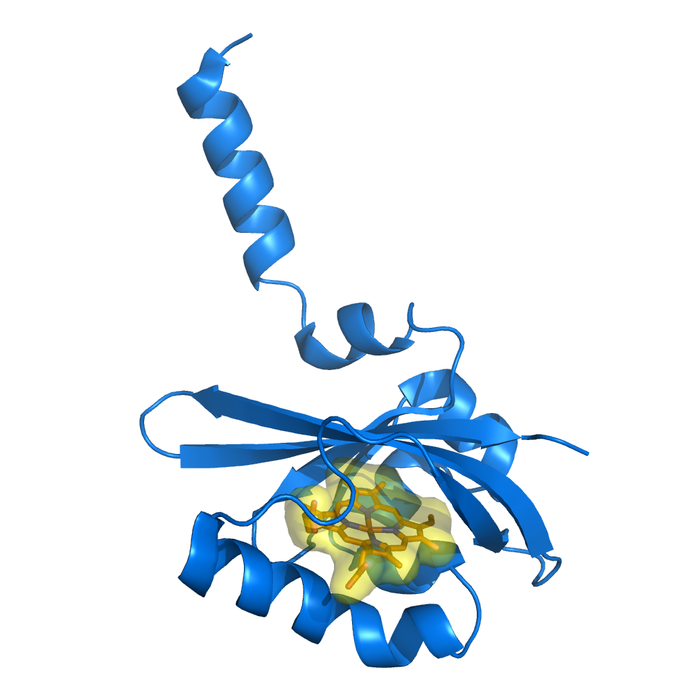
院内感染菌のヘムトランスポーター BhuUV-T
- Naoe, Y., Nakamura, N., Doi, A., Sawabe, M., Nakamura, H., Shiro, Y., and Sugimoto, H. "Crystal structure of bacterial haem importer complex in the inward-facing conformation. Nature Commun. 13411 (2016) [doi]
- 病原菌が鉄を細胞内に取り込む仕組み-細胞膜で働くヘム輸送体タンパク質の立体構造を解明- プレスリリース (2016年11月10日)
|
| 
|
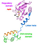
ジフテリア菌のヘムシグナル伝達因子(レスポンスレギュレータ)
- Doi, A., Nakamura, H., Shiro, Y., and Sugimoto, H. "Structure of the response regulator ChrA in the haem-sensing two-component system of Corynebacterium diphtheriae". Acta Crystallogr. F Struct. Biol. Commun. 71, 966-971 (2015).[PubMed]
|
| 
|
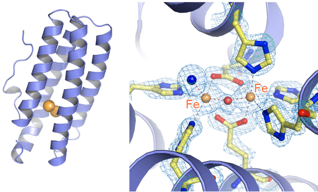
二核鉄サイトを改変したヘムエリスリン (大阪大学との共同研究)
- Okamoto, Y., Onoda, A., Sugimoto, H., Takano, Y., Hirota, S., Kurtz, D. M., Shiro, Y., Hayashi, T. "H2O2-dependent substrate oxidation by an engineered diiron site in a bacterial hemerythrin". Chem. Commun. 50, 3421-3423 (2014)
- Okamoto, Y., Onoda, A., Sugimoto, H., Takano, Y., Hirota, S., Kurtz, D. M., Jr., Shiro, Y., Hayashi, T. "Crystal Structure, Exogenous Ligand Binding, and Redox Properties of an Engineered Diiron Active Site in a Bacterial Hemerythrin". Inorg. Chem. 52, 13014-13020 (2013)
|
| 
|
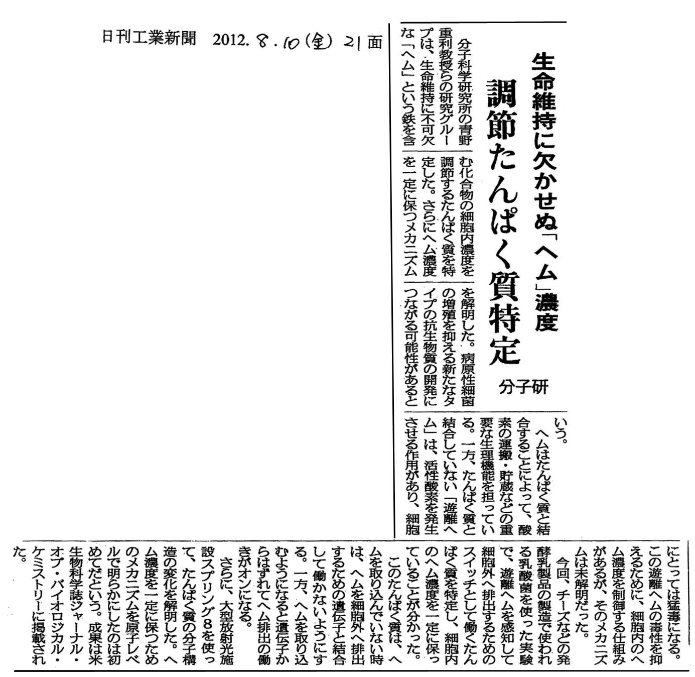
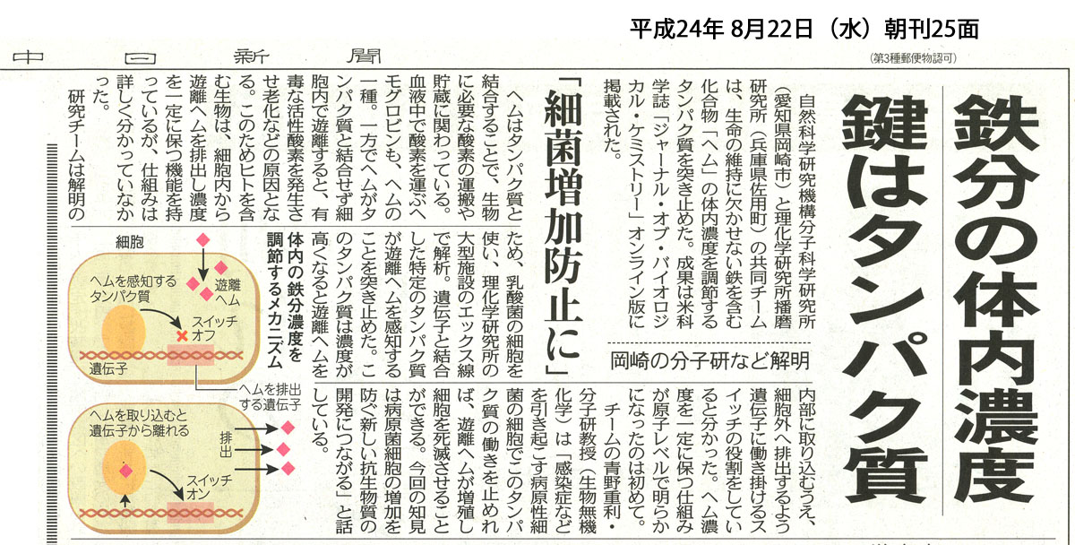
細胞内ヘムセンサータンパク質 HrtR (分子研との共同研究)
- Sawai, H., Yamanaka, M., Sugimoto, H., Shiro, Y., Aono, S. "Structural basis for the transcriptional regulation of heme homeostasis in Lactococcus lactis." J. Biol. Chem 287, 30755-30768 (2012) [PubMed]
|
| 
|
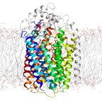
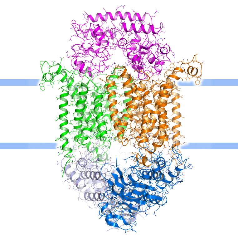
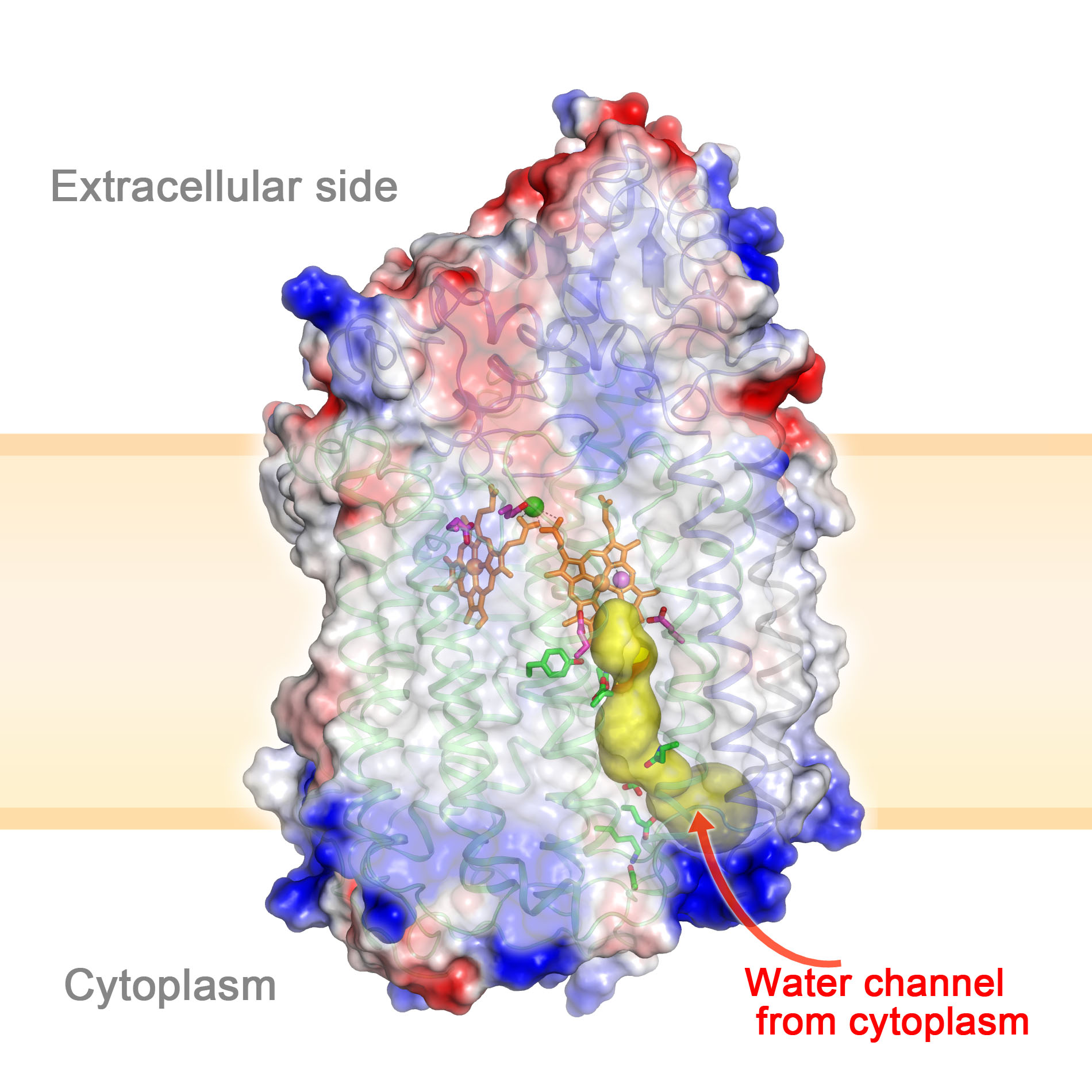
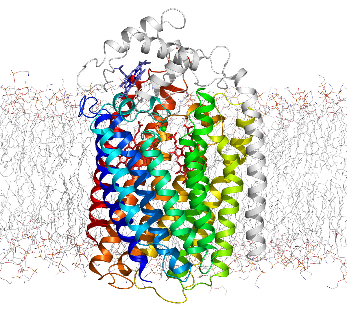
キノール依存型一酸化窒素還元酵素 qNOR
- Matsumoto, Y., Tosha, T., Pisliakov, A. V., Hino, T., Sugimoto, H., Nagano, S., Sugita, Y., Shiro, Y. "Crystal structure of quinol-dependent nitric oxide reductase from Geobacillus stearothermophilus." Nature Struct. Mol. Biol. 19, 238-245 (2012) [PubMed]
|
| 
|
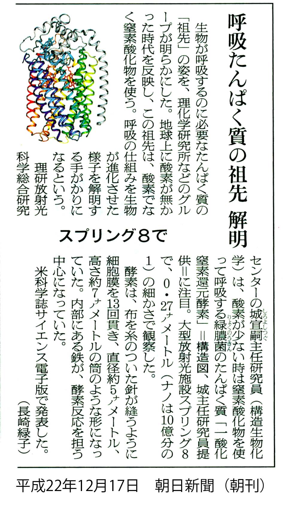
一酸化窒素還元酵素 cNOR (嫌気呼吸から酸素呼吸への進化のしくみ)
- Hino, T., Matsumoto, Y., Nagano, S., Sugimoto, H., Y., F., Murata, T., Iwata, S., Shiro, Y. Structural basis of biological N2O generation by bacterial nitric oxide reductase. Science 330, 1666-1670 (2010) [PubMed]
|
| 
|
アルドキシムからニトリルを合成する酵素 OxdRE
- Sawai, H., Sugimoto, H., Kato, Y., Asano, Y., Shiro, Y., Aono, S. X-ray crystal
structure of the michaelis complex of aldoxime dehydratase. J. Biol.
Chem. 284, 32089-96 (2009)
[PubMed]
|
| 
|
ヒスチジンキナーゼ・レスポンスレギュレーター複合体
- Yamada, S., Sugimoto, H., Kobayashi, M., Ohno, A., Nakamura, H., Shiro, Y. Structure of
PAS-linked histidine kinase and the response regulator complex. Structure
17, 1333-1344 (2009)
[PubMed]
- Seeing the sencein
it all (RIKEN Research Highlights) 25 Dec. 2009
-
世界初の構造を解明 --微生物の環境適応センサータンパク質-- (サイエンスニュースオンデマンド) – 2009年11月2日
-
微生物の環境適応センサータンパク質の構造を解明 --光、熱、酸素、ストレスなどの環境変化への応答機構を発見--
(プレスリリース) 2009年10月14日
|
| 
|
シアン色の蛍光タンパク質
- Kikuchi, A.,
Fukumura, E., Karasawa, S., Shiro, Y., Miyawaki, A. Crystal structure of a
new cyan fluorescent protein and its hue-shifted variants. Biochemistry
48, 5276-5283 (2009)
[PubMed]
|
| 
|
ビタミンD 活性化酵素CYP105A1
- Sugimoto, H., Shinkyo, R., Hayashi, K., Yoneda, S.,
Yamada, M., Kamakura, M., Ikushiro, S., Shiro, Y., Sakaki, T. Crystal
structure of CYP105A1 (P450SU-1) in complex with 1alpha,25-dihydroxyvitamin D3.
Biochemistry 47, 4017-4027 (2008)
- Hayashi, K., Sugimoto, H., Shinkyo, R., Yamada, M.,
Ikeda, S., Ikushiro, S., Kamakura, M., Shiro, Y., Sakaki, T. Structure-based
design of a highly active vitamin D hydroxylase from Streptomyces griseolus
CYP105A1. Biochemistry 47, 11964-11972 (2008)
|
| 
|
オレンジ色の蛍光タンパク質(クサビラオレンジ)
- Kikuchi, A.,
Fukumura, E., Karasawa, S., Mizuno, H., Miyawaki, A., Shiro, Y. Structural
characterization of a thiazoline-containing chromophore in an orange
fluorescent protein, monomeric Kusabira Orange. Biochemistry 47,
11573-11580 (2008)
[PubMed]
|
| 
|
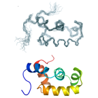
ドロンパ
- Mizuno, H., Mal, T. K., Walchli, M., Kikuchi, A.,
Fukano, T., Ando, R., Jeyakanthan, J., Taka, J., Shiro, Y., Ikura, M.,
Miyawaki, A. Light-dependent regulation of structural flexibility in a
photochromic fluorescent protein. Proc. Natl. Acad. Sci. USA 105,
9227-9232 (2008)
-
蛍光タンパク質「ドロンパ」のフォトクロミズムの分子機構を解明へ –X線結晶構造解析と核磁気共鳴(NMR)を駆使し、
ドロンパの動的構造を決定—(プレスリリース)
|
|
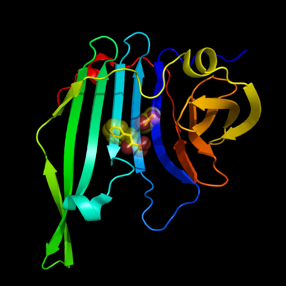
微生物色素ビオラセイン合成タンパク質 VioE
- Hirano, S., Asamizu, S., Onaka, H., Shiro, Y., Nagano, S. Crystal Structure of VioE,
a Key Player in the Construction of the Molecular Skeleton of Violacein. J.
Biol. Chem. 283, 6459-6466 (2008)
|
| 
|
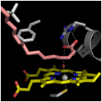
人工修飾ヘムをもつミオグロビン(片足ヘムに置換)
- Harada, K., Makino, M., Sugimoto, H., Hirota, S., Matsuo, T., Shiro, Y., Hisaeda, Y.,
Hayashi, T. Structure and ligand binding properties of myoglobins
reconstituted with monodepropionated heme: functional role of each heme
propionate side chain. Biochemistry 46, 9406-9416 (2007)
|
|
|
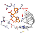
抗がん剤インドロカルバゾールの骨組みを構築する酵素 P450StaP
- Makino, M., Sugimoto, H., Shiro, Y., Asamizu, S.,
Onaka, H., Nagano, S. Crystal structures and catalytic mechanism of
cytochrome P450 StaP that produces the indolocarbazole skeleton. Proc.
Natl. Acad. Sci. USA 104, 11591-11596 (2007)
- Wang, Y., Chen, H., Makino, M., Shiro, Y., Nagano, S.,
Asamizu, S., Onaka, H., Shaik, S. Theoretical and experimental studies of the
conversion of chromopyrrolic acid to an antitumor derivative by cytochrome
P450 StaP: the catalytic role of water molecules. J. Am. Chem. Soc.
131, 6748-6762 (2009)
-
抗がん剤インドロカルバゾールの骨組みを構築する酵素の立体構造を解明
--放線菌がインドロカルバゾールを創り出すメカニズムの一端が明らかに--プレスリリース
-
Core structures -- RIKEN Research Highlights (14 Sep 2007)
-
Water-powered reactions -- RIKEN Research Highlights (26 Jun 2009)
|
|
|
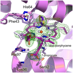
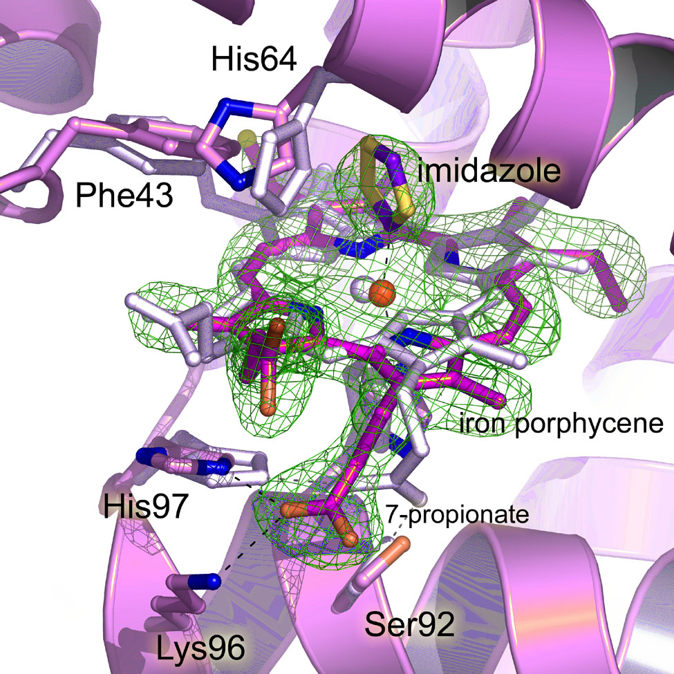
ポルフィセン置換ミオグロビン
- Hayashi, T., Murata, D., Makino, M., Sugimoto, H., Matsuo, T., Sato, H., Shiro, Y.,
Hisaeda, Y. Crystal structure and peroxidase activity of myoglobin
reconstituted with iron porphycene. Inorg. Chem. 45,
10530-10536 (2006)
|
| 
|
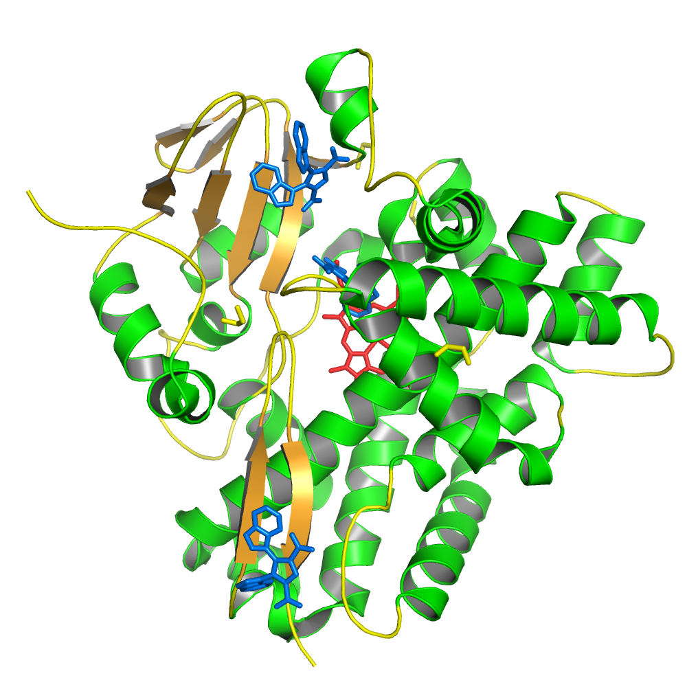
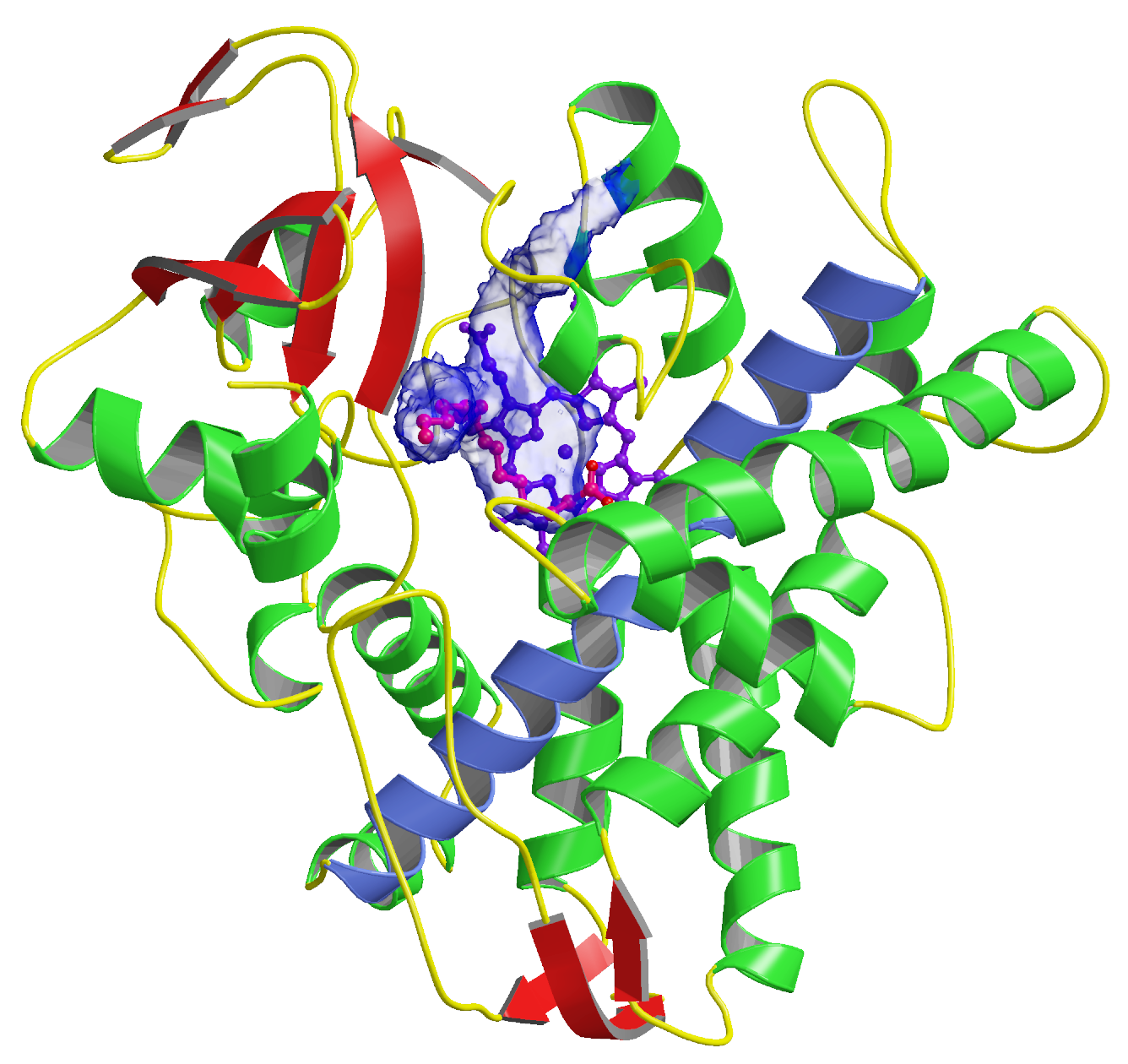
インドールアミン 2,3-ジオキシゲナーゼ (IDO)
- Sugimoto, H., Oda, S., Otsuki, T., Hino, T., Yoshida,
T., Shiro, Y. Crystal structure of human indoleamine 2,3-dioxygenase:
catalytic mechanism of O2 incorporation by a heme-containing dioxygenase. Proc.
Natl. Acad. Sci. USA 103, 2611-2616 (2006)
- Chung, L. W., Li, X., Sugimoto, H., Shiro, Y.,
Morokuma, K. Density functional theory study on a missing piece in
understanding of heme chemistry: the reaction mechanism for indoleamine
2,3-dioxygenase and tryptophan 2,3-dioxygenase. J. Am. Chem. Soc.
130, 12299-12309 (2008)
-
発見から50年、酸素添加酵素「ジオキシゲナーゼ」の反応機構が明らかに --プレスリリース
-
Missing piece gets a work over -- RIKENリサーチハイライト 2009年1月16日
|
| 
|
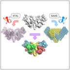
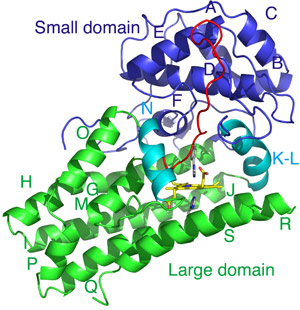
X線結晶解析と線小角散乱法で視るヒスチジンキナーゼの構造
- Yamada, S., Akiyama, S., Sugimoto, H., Kumita, H., Ito,
K., Fujisawa, T., Nakamura, H., Shiro, Y. The signaling pathway in histidine
kinase and the response regulator complex revealed by X-ray crystallography
and solution scattering. J. Mol. Biol. 362, 123-139 (2006)
|
| 
|
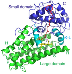
ヒト第4のグロビン:サイトグロビン (Cgb)
- Sugimoto, H., Makino, M., Sawai, H., Kawada, N.,
Yoshizato, K., Shiro, Y. Structural basis of human cytoglobin for ligand binding.
J. Mol. Biol. 339, 873-885 (2004)
- Makino, M., Sugimoto, H., Sawai, H., Kawada, N.,
Yoshizato, K., Shiro, Y. High-resolution structure of human cytoglobin:
identification of extra N- and C-termini and a new dimerization mode. Acta
Crystallogr. Sect. D Biol. Crystallogr. 62, 671-677
(2006)
|
| 
|
転写活性因子FixJのDNA結合ドメイン
- Kurashima-Ito, K., Kasai, Y., Hosono, K., Tamura, K.,
Oue, S., Isogai, M., Ito, Y., Nakamura, H., Shiro, Y. Solution structure of
the C-terminal transcriptional activator domain of FixJ from Sinorhizobium
meliloti and its recognition of the fixK promoter. Biochemistry
44, 14835-14844 (2005)
|
| 
|
脂肪酸代謝酵素P450BSbeta
- Lee, D. S., Yamada, A., Sugimoto, H., Matsunaga, I.,
Ogura, H., Ichihara, K., Adachi, S., Park, S. Y., Shiro, Y. Substrate
recognition and molecular mechanism of fatty acid hydroxylation by cytochrome
P450 from Bacillus subtilis. Crystallographic, spectroscopic, and
mutational studies. J. Biol. Chem. 278, 9761-9767 (2003)
- Shoji, O., Fujishiro, T., Nakajima, H., Kim, M.,
Nagano, S., Shiro, Y., Watanabe, Y. Hydrogen peroxide dependent
monooxygenations by tricking the substrate recognition of cytochrome
P450BSbeta. Angew. Chem. Int. Ed. 46, 3656-3659 (2007)
|
|
|
ヘモグロビンの反応中間状態
- Adachi, S., Park, S. Y., Tame, J. R., Shiro, Y., Shibayama, N. Direct observation of
photolysis-induced tertiary structural changes in hemoglobin. Proc.
Natl. Acad. Sci. USA 100, 7039-7044 (2003)
-
ヘモグロビンへの配位子結合過程を直接観測 -- プレスリリース(H15.6.9)
|
|
ビリベルジン還元酵素
- Kikuchi, A., Park, S. Y., Miyatake, H., Sun, D., Sato, M., Yoshida, T., Shiro, Y. Crystal
structure of rat biliverdin reductase. Nat. Struct. Biol. 8,
221-225 (2001)
-
金属タンパク質の構造と機能の「専門書」をつくる -- 理研ニュース 2001年7月号
|
|
|
酸素センサータンパク質 FixLのPASドメイン
- Miyatake, H., Mukai, M., Park, S. Y., Adachi, S., Tamura, K., Nakamura, H., Nakamura, K.,
Tsuchiya, T., Iizuka, T., Shiro, Y. Sensory mechanism of oxygen sensor FixL
from Rhizobium meliloti: crystallographic, mutagenesis and resonance
Raman spectroscopic studies. J. Mol. Biol. 301,
415-431 (2000)
|
|
高熱性細菌のP450 (CYP1119)
- Denisov, I. G., Hung, S. C., Weiss, K. E., McLean, M. A., Shiro, Y., Park, S. Y.,
Champion, P. M., Sligar, S. G. Characterization of the oxygenated intermediate
of the thermophilic cytochrome P450 CYP119. J. Inorg. Biochem. 87,
215-226 (2001)
|
|
|
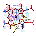
カビの一酸化窒素還元酵素 P450nor
- Park, S.Y., Shimizu, H., Adachi, S., Nakagawa, A., Tanaka, I., Nakahara, K., Shoun, H.,
Obayashi, E., Nakamura, H., Iizuka, T., Shiro, Y. Crystal structure of nitric
oxide reductase from denitrifying fungus Fusarium oxysporum. Nature
truct. Biol. 4, 827-32 (1997)
- Shimizu, H., Park, S. Y., Shiro, Y., Adachi, S. X-ray structure of nitric oxide reductase
(cytochrome P450nor) at atomic resolution. Acta Crystallogr. Sect. D
Biol. Crystallogr. 58, 81-89 (2002)
|
