Dec. 21, 2018 Research Highlight Chemistry
Extra genetic letters cause target DNA to light up
A novel test detects pathogens or other target organisms by lighting up their DNA using an expanded genetic alphabet
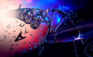 Figure 1: By adding two more bases to the genetic alphabet, RIKEN researchers have developed a naked-eye detection technique for identifying target DNA. © IAN CUMING/IKON IMAGES/SCIENCE PHOTO LIBRARY
Tiny traces of DNA produced by drug-resistant superbugs or other target organisms can now be spotted with the naked eye, thanks to a technique developed by researchers at RIKEN and A*STAR1. It could be used to identify target DNA and diagnose diseases using simple instruments.
DNA consists of long strands of chemical ‘letters’, or bases, that make up the genetic code. Nature uses just four bases: adenine (A), cytosine (C), guanine (G) and thymine (T). But Ichiro Hirao, who did the work at the RIKEN Center for Life Science Technologies before joining the A*STAR Institute of Bioengineering and Nanotechnology (IBN) in Singapore, has been seeking to expand the genetic alphabet to six by creating a new pair of bases: Ds and Px.
Hirao’s team has now shown that these extra bases can be used to unmask the presence of target DNA. They achieved this by amplifying the target DNA while simultaneously incorporating tags that emit visible light when illuminated by an ultraviolet flashlight.
The researchers added their bases to a commonly used technique known as the polymerase chain reaction (PCR), which uses a trace of DNA as a template to make lots of identical copies for analysis. One example of its many uses is in forensics, where it is used to amplify DNA traces found at crime scenes.
Hirao and his team made short strips of DNA called PCR primers, which contained several non-natural DNA bases. A pair of light-capturing ‘s’ bases were included, alongside the Ds base. “These primers can be easily prepared by conventional chemical synthesis,” notes Hirao.
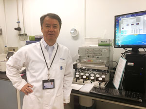 Ichiro Hirao in the laboratory. © 2018 RIKEN
The primer was then added to the PCR mixture, along with free Px nucleobases to which the researchers had attached an orange-light-emitting Cy3 side chain. In the presence of the target DNA—from a drug-resistant bacterium, for example—new copies of double-stranded DNA form. Ds pairs with Px in the new copies, which brings the Cy3 group close to the two s bases. When ultraviolet light is shone on the sample, energy captured by the s bases passes to the Cy3 group, and the sample glows orange (Fig. 1). If the target DNA is not present, the s bases and the Cy3 groups remain dissociated and no light is produced.
Another A*STAR team has recently developed a miniaturized microfluidic device for portable PCR. “By applying our technique to the microfluidics chip, we hope to develop a simple and small detection–diagnostic system that can be used in the field,” Hirao comments.
Related contents
- A tighter fit with artificial DNA
- Multifunctional molecules aid mutation analysis
- Highly efficient molecular probe developed for real-time PCR monitoring
References
- 1. Yamashige, R., Kimoto, M., Okumura, R. & Hirao, I. Visual detection of amplified DNA by polymerase chain reaction using a genetic alphabet expansion system. Journal of the American Chemical Society 140, 14038–14041 (2018). doi: 10.1021/jacs.8b08121