Feb. 21, 2008 Research Highlight Biology
Neural architecture
The neurons in the primary visual cortex processing high- and low-frequency images are distinct
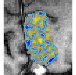 Figure 1: The pattern of temporal-frequency domains in the human primary visual cortex shows distinct characteristics. Low temporal-frequency domains appear to be continuous (yellow) whereas the high temporal-frequency domains are more isolated (blue). © Nature Neuroscience / Nature Publishing Group / 10 / 1406 (2007)
Neuroscientists from the RIKEN Brain Science Institute, Wako, and New York University have used functional magnetic resonance imaging (fMRI) to study the organization of neurons in the primary visual cortex (V1) of humans and establish that the temporal frequency of a stimulus activates specific V1 neurons.
The V1 is an area at the back of the brain where the first stage of visual processing takes place. Although this is one of the most heavily studied parts of the visual cortex, little is known about how its neurons are arranged. In general, neurons with similar selectivity for visual stimuli cluster together. For example, V1 neurons that process stimuli from each eye are grouped into pillars, called ocular dominance columns.
V1 neurons are highly sensitive to the contrast, orientation, and spatio-temporal frequency of a visual stimulus. Temporal frequency is an important determinant of how moving images are processed by the brain and is a measure of how often an image appears in the visual field. This attribute is also of particular interest to RIKEN researcher Pei Sun and his team, headed by Keiji Tanaka and Kang Cheng, who have determined that images appearing less frequently over time are handled by neurons that arrange themselves differently to those that are activated by more frequently appearing images.
The fMRI technique allows the function and anatomical structure of the brain to be studied live and works by measuring the level of oxygen in the blood immediately after a neuron has been active, giving a pattern of which neurons have been triggered by a stimulus.
The team has shown that separate domains in human V1 respond preferentially to low- and high-temporal frequencies. The former appear to be continuous, whereas the latter seem to be more like isolated islands with no particular orientation (Fig. 1).
This study provides direct physiological evidence that different temporal frequencies are preferentially processed by spatially segregated streams in human V1. The work recently published in Nature Neuroscience1 is the first to show neuronal organization specific to temporal frequency in primate V1.
Evidence of these separate neural regions will assist further study into human perception of moving images and help to develop a map of the neural architecture of the brain. Pei plans to develop the fMRI technique as “it could link animal and human behavioral studies, giving a better picture of how information is processed by the brain,” he says.
References
- 1. Sun, P., Ueno, K., Waggoner, R.A., Gardner, J.L., Tanaka, K. & Cheng, K. A temporal frequency-dependent functional architecture in human V1 revealed by high-resolution fMRI. Nature Neuroscience 10, 1404–1406 (2007). doi: 10.1038/nn1983