Oct. 16, 2008 Research Highlight Biology
Guiding the decision-making process
Identification of a novel protein involved in embryonic development leads to new insights into the first stage of neural development
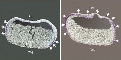 Figure 1: Xenopus embryos stained to indicate expression of a mesodermal marker. In the left panel, an unmodified embryo exhibits the marker in the region between the ectodermal animal pole region (An) and the vegetal pole (Veg), where the endoderm is formed. The embryo in the right panel has greatly reduced XFDL production, resulting in the formation of mesoderm in a typically ectodermal region of the embryo. Reproduced, with permission, from Ref. 1 © 2008 Elsevier
In developing animal embryos, stem cells soon differentiate into three distinct layers of tissue, the primary germ layers. These are the endoderm, mesoderm and ectoderm, and each subsequently develops into a specific subset of tissues and organs. These differentiating cells follow a highly complex ‘decision-making’ process, and a lot of ambiguity still remains as to why, for example, an ectodermal cell became an ectodermal cell.
Yoshiki Sasai of the RIKEN Center for Developmental Biology in Kobe views the exploration of this process as a long-term mission. “Fifteen years ago, I isolated the neural inducer chordin, which induces neural progenitors from uncommitted ectoderm,” he says. “However, how the uncommitted ectoderm develops from pluripotent cells has remained unknown—so it has been my 15-year-old homework to elucidate it!”
In the frog species Xenopus laevis—a popular animal model for developmental studies—germ-layer differentiation is triggered via two sets of signals: some derived from maternally produced factors, and others generated by the zygote itself. Previous research has identified several candidate maternal factors, but the mechanisms involved in the latter pathway have proven more elusive, and Sasai’s team has focused much of their recent effort on finding zygotic factors potentially responsible for ectoderm formation.
In their most recent study, Sasai’s team identified a previously uncharacterized protein, XFDL, which exhibits a marked ability to block mesoderm formation1 (Fig. 1). Elevated XFDL levels lead to inhibition of mesoderm-specific genes, while reduced levels of the protein lead to broader expression of these genes in embryonic regions that normally form ectoderm.
Subsequent experiments revealed that XFDL acts by interacting with p53, a transcription-regulating protein with a known role in mesoderm formation, and directly interfering with its ability to bind to DNA and activate its target genes. When XFDL was prevented from interacting with p53, it lost its ability to regulate ectoderm differentiation—an unexpected finding, according to Sasai. “Although p53 is implicated in the regulation of mesodermal development, we did not think it had such a profound function in the binary decision of ectodermal versus mesodermal determination,” he says.
The researchers have identified two XFDL-related proteins in mice, both of which also inhibit mesoderm formation in Xenopus, indicating that mammalian germ layer formation may also be regulated via a similar pathway—a possibility that Sasai’s team is currently exploring more closely. “Our preliminary studies suggest that this is the case at least with in vitro differentiation of mammalian embryonic stem cells,” he says.
References
- 1. Sasai, N., Yakura, R., Kamiya, D., Nakazawa, Y. & Sasai, Y. Ectodermal factor restricts mesoderm differentiation by inhibiting p53. Cell 133, 878–890 (2008). doi: 10.1016/j.cell.2008.03.035