Oct. 23, 2009 Research Highlight Biology
Helping neurons find their way
Variations in the spatial distribution of cellular signaling molecules provide the information needed to steer neuron growth within the brain
As the brain develops, neuronal axons extend outward in search of other neurons, all the while receiving ‘directions’ from the extracellular environment in the form of chemical signals that indicate when and where these growing axons should turn. For example, axons exposed to a gradient distribution of nerve growth factor (NGF) protein will automatically steer in the direction of highest NGF concentration.
“NGF is one of the most extensively studied molecules that direct axon elongation,” explains Hiroyuki Kamiguchi of the RIKEN Brain Science Institute in Wako. “However, it has remained unclear for a long time how axons change the direction of elongation in response to NGF.”
NGF-mediated turning is facilitated in part by the cellular signaling molecule inositol trisphosphate (IP3), which in turn governs the intracellular release of calcium ions—an essential component of NGF’s chemo-attractive action. By applying advanced methods for molecular-resolution live cell imaging, Kamiguchi and his colleagues have now gained valuable insights into how this process directs axonal guidance1.
The researchers cultured chick-derived neurons expressing a genetically encoded sensor that fluoresces at specific wavelengths in the presence of IP3, and then observed how individual neurons responded to an NGF gradient in the vicinity of the growth cone—the leading edge of a growing axon. They immediately noted the establishment of an asymmetric distribution of IP3 within the growth cone and an elevated signal on the growth cone side exposed to higher NGF levels; this is mirrored by a similarly uneven distribution of IP3-induced calcium release. This asymmetry correlates directly with axonal turning such that the growth cone steers in the direction established by the highest levels of NGF, IP3 and calcium ion (Ca2+) release (Fig. 1).
The development of techniques for accurately detecting potentially subtle variations in IP3 distribution was a key component of their success in this work. “We needed to detect 1% differences in fluorescence emission from the IP3 sensor between both sides of the growth cone,” says Kamiguchi.
However, he considers even the mere existence of such a gradient across the 10–20 micron width of the growth cone to be fairly surprising. “Because IP3 diffuses so rapidly in cytoplasm, it has not been viewed as a highly localized messenger,” he says. “This suggests the existence of robust degradation machinery to localize IP3 signals to one side of the growth cone.”
These insights into how neurons establish direction-specific signaling profiles should provide helpful starting points for understanding other models of cell polarization and migration.
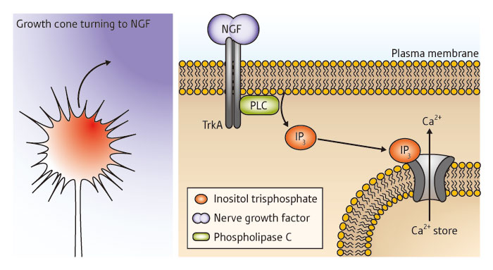 Figure 1: Schematic showing an axonal growth cone being guided in the direction of highest concentration of NGF (purple) via the establishment of an asymmetric internal gradient of IP3 (red) (left). This action is mediated by elevated NGF receptor (TrkA) binding at the axonal surface, resulting in increased IP3 production via the enzyme phospholipase C (PLC); this subsequently induces release of Ca2+, which stimulates axonal turning (right).
References
- 1. Akiyama, H., Matsu-ura, T., Mikoshiba, K. & Kamiguchi, H. Control of neuronal growth cone navigation by asymmetric inositol 1,4,5-trisphosphate signals. Science Signaling 2, ra34 (2009). doi: 10.1126/scisignal.2000196