Aug. 5, 2011 Research Highlight Biology
Unraveling genomic changes in the brain
Differences in the chemical modification of DNA within neurons from different individuals may hint at new ways of understanding the roots of mental illness
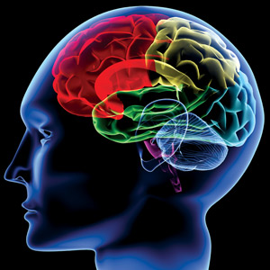 Figure 1: The prefrontal cortex is at the front of the cerebrum (in red) of the brain. © 2011 iStockphoto/ Yakobchuk
Scientists have known for some time that individual organisms are far more than the sum of their gene sequences. So-called epigenetic variations encompass a diverse array of chemical modifications to DNA that leave the core nucleotide sequence unchanged, but can nevertheless exert powerful effects on gene expression behavior.
A modification commonly associated with the silencing of gene activity in mammals, for example, is the enzyme-mediated attachment of a methyl group onto a cytosine base within a ‘CpG dinucleotide’, a segment of chromosomal DNA in which cytosine is immediately followed by guanine. Different genes are subject to such methylation in particular tissues, but methylation can also differ markedly in the same tissues between individuals, with potentially profound functional consequences.
According to Tadafumi Kato of the RIKEN Brain Science Institute in Wako, abnormalities in DNA methylation within the brain could even play a role in a variety of neuropsychiatric conditions, such as bipolar disorder. “Mental disorders are the result of gene–environment interaction,” he explains, “and DNA methylation may be affected by early environmental effects.”
As a first step toward testing this hypothesis, Kato’s group teamed up with The University of Tokyo researcher Kazuya Iwamoto, along with colleagues in Japan and the United States, to survey genome-wide methylation in the brains of different individuals1. For their analysis, they examined postmortem tissue from the prefrontal cortex of the cerebrum (Fig. 1), a region of the brain with a leading role in decision-making behavior.
The nature of neurons
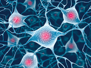 Figure 2: Neurons separated from the brain were profiled for methylation, which can vary greatly between individuals. © 2011 iStockphoto/ ktsimage
Kato and colleagues used the neuron-specific protein NeuN as a marker for separating out actual neurons from the larger population of glial cells (Fig. 2), which play a vital part in supporting neuronal function. “If we extracted DNA from the bulk cerebral cortex, the DNA methylation data would mainly reflect the glial cells,” says Kato. “Thus, we need to separate out the neurons from the brain.”
This approach enabled the researchers to perform a direct comparison of the two cell classes. Indeed, their initial assessment confirmed that the methylation profile of the NeuN-negative (glial) cells displayed a significantly greater degree of similarity to that of bulk cortical samples relative to the NeuN-positive (neuronal) subpopulation. In general, bulk cortical preparations and glial cell fractions also exhibited notably higher levels of overall genomic methylation in comparison to neuronal fractions.
Kato, Iwamoto and colleagues used a platform known as a ‘tiling array’ to precisely map the positions of these methylation sites within the genome. Based on these data, they were able to compile collections of methylated regions (MRs) that were specifically neuronal, non-neuronal or common to both cell populations. Many of the MRs were located within promoters, which are stretches of DNA that directly control the activity of nearby genes. The researchers also identified hundreds of genes that display similar patterns of epigenetic modification within each of the three categories. Intriguingly, they noted that promoters that were specifically methylated within the glial population regulate a number of established neuronal genes, encoding factors associated with ion transport and neurotransmitter signaling.
In a prior study, the researchers had investigated overall gene expression within the brain, identifying patterns of transcriptional regulation that enabled them to cluster various genes into discreet functional ‘modules’2. Kato and colleagues used these findings to determine whether any of these modules were particularly enriched among any of the subsets of methylated genes.
Several of the genes that appeared to be specifically methylated, and therefore silenced, among non-neuronal cells belonged to a module associated with mitochondrial function. The activity of these organelles is known to be considerably enriched in neurons. Conversely, the researchers found evidence that silencing among neuronal populations appears to affect genes associated with the function of astrocytes, a glial subpopulation.
Defining our differences
In a comparison of the distribution of MRs between different individuals, Kato and colleagues were surprised to find a very high level of variability in neuronal methylation from person to person. For any given sample, the proportion of unique MRs was significantly higher in genetic material from neuronal cells relative to non-neuronal cells, while the proportion of shared MRs was significantly lower. These results were subsequently mirrored in an independent assessment of 24 additional brain tissue samples.
“Our brain function is continuously changing, and we all have different brain functional status relative to each other, so it seems reasonable that DNA methylation status is more flexible in neurons than in glial cells,” says Kato. “However, we did not anticipate this finding before starting our project.”
The researchers note that this remains a broad survey of genomic modification, especially for an organ as complex as the brain. The cortex alone contains numerous subtypes of neurons, each of which may manifest its own particular profile of methylation; likewise, relative populations of different neuronal types could differ significantly from person to person. In addition, the method applied in this study is not suitable for tracking every instance of methylation; for example, it is incapable of detecting hydroxymethyl cytosine nucleotides, although such modifications are believed to be widespread in both mouse and human brains.
Nevertheless, the researchers’ work represents one of the most detailed epigenetic analyses of the brain to date, and the resulting data have encouraged Kato and his colleagues to dig deeper in an effort to untangle potential links between variable methylation and mental health. “We are now studying the DNA methylation status of neuronal nuclei in the post-mortem brains of patients with bipolar disorder and schizophrenia,” says Kato.
References
- 1. Iwamoto, K., Bundo, M., Ueda, J., Oldham, M.C., Ukai, W., Hashimoto, E., Saito, T., Geschwind, D.H. & Kato, T. Neurons show distinctive DNA methylatinon profile and higher interindividual variations compared with non-neurons. Genome Research 21, 688–696 (2011). doi: 10.1101/gr.112755.110
- 2. Oldham, M.C., Konopka, G., Iwamoto, K., Langfelder, P., Kato, T., Horvath, S. & Geschwind, D.H. Functional organization of the transcriptome in human brain. Nature Neuroscience 11, 1271–1282 (2008). doi: 10.1038/nn.2207
About the Researcher
Tadafumi Kato

Tadafumi Kato was born in Tokyo, Japan, in 1963. He graduated from the Faculty of Medicine, The University of Tokyo, in 1988. After residency training at The University of Tokyo Hospital, he obtained his PhD in 1995 from Shiga University of Medical Science. After working as a lecturer at the Department of Neuropsychiatry, The University of Tokyo, he was appointed as a team leader at the RIKEN Brain Science Institute in 2001. His research focuses on the neurobiology of bipolar disorder.