Nov. 4, 2011 Research Highlight Biology
Shaping up for cell division
Preparing chromosomes for cell division is a balancing act involving a tug-of-war between opposing molecular actions
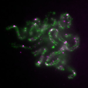 Figure 1: Mitotic chromosomes assembled in the Xenopuscell-free system. Condensin I (green) and II (magenta) display distinct localizations within the chromosomes. © 2011 Tatsuya Hirano
The shape of chromosomes is determined by the relative levels of key protein complexes, research conducted by Keishi Shintomi and Tatsuya Hirano of the RIKEN Advanced Science Institute has shown1.
As a cell prepares to divide via the process called mitosis, chromatin—the material in which DNA is packaged—condenses to form discrete rod-shaped structures called chromosomes. Each chromosome contains duplicated chromatids—sister chromatids—that are aligned in parallel. After ‘mitotic chromosome condensation’ is complete, the paired chromatids segregate such that each daughter cell receives one of each pair.
“For well over a century, biologists have noticed that the shape of condensed chromosomes is highly characteristic, but varies among different organisms or among different developmental stages in a single organism,” explains Hirano. “We are interested in understanding how the shape of chromosomes is determined at a molecular level.”
Hirano’s group previously discovered that mitotic chromosome condensation requires the action of two protein complexes, known as condensins I and II. This group and others have shown that a third protein complex called cohesin is responsible for the pairing of sister chromatids within a chromosome.
To test exactly how condensins and cohesin may contribute to shaping of chromosomes, Shintomi and Hirano turned to a cell-free system based on extracts prepared from the eggs of the frog Xenopus laevis. “The Xenopus system perfectly suited our purposes because it enables us to recapitulate many chromosomal events, including chromosome condensation, in a test tube in a cell-cycle regulated manner (Fig. 1),” says Hirano.
To achieve their goal, the researchers then had to develop a series of sophisticated experimental protocols to precisely manipulate the levels of condensins I and II and cohesin present in the extracts.
Under the standard condition, chromosomes assembled in this cell-free system tended to be long and thin, which are general characteristics of chromosomes observed in early embryos. Strikingly, however, when the ratio of condensin I to II was reduced, they became shorter and thicker, being reminiscent of chromosomes observed in later stages of development. Further experiments revealed that cohesin works with condensin I and counteracts condensin II to properly place sister chromatids within a chromosome. Thus, their actions can be likened to a molecular ‘tug-of-war’.
“Our findings demonstrated that chromosome shape is achieved by an exquisite balance between condensin I and II and cohesin,” says Hirano. “Such a concept had been suspected for a long time, but has never been demonstrated so beautifully and convincingly until now.”
References
- 1. Shintomi, K. & Hirano, T. The relative ratio of condensin I to II determines chromosome shapes. Genes & Development 25, 1464–1469 (2011). doi: 10.1101/gad.2060311