Dec. 20, 2013 Research Highlight Biology
Mapping objects in the brain
A brain region that responds to a particular category of objects is found to consist of small clusters of neurons encoding visual features of these objects
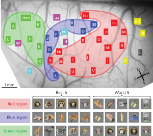 Figure 1: Neuronal activity during exposure to various images reveals distinct spatial groupings. The red region, for example, responds well to face stimuli. © 2013 Takayuki Sato, RIKEN Brain Science Institute
The ability to recognize objects in the environment is mediated by the brain’s ability to integrate and process massive amounts of visual information. A research group led by Takayuki Sato and Manabu Tanifuji from the RIKEN Brain Science Institute has now discovered that in macaque monkeys, this remarkable ability is supported by mosaic-like structures in the anterior inferior temporal (IT) cortex, where localized clusters of neurons encode different visual features in an organized hierarchy1.
Two competing models have been proposed to explain the functional organization of brain regions that underlies object recognition in primates. One model states that discrete brain ‘modules’ process stimuli from particular categories, such as faces, with object recognition arising from communication among the modules. The other model postulates that the visual cortex extracts generic features, which are then composited to recognize specific objects. Since both models are based on measurements of functional signals produced by metabolic changes associated with neural activity rather than measurements of the neuronal activity itself, the precise underlying mechanism responsible for object recognition has remained unclear.
To resolve this debate, the researchers undertook dense electrophysiological mapping of neural activity in anesthetized macaque monkeys exposed to a series of color images from different object categories: faces, hands, bodies, food and various other objects. Sato and his colleagues directly recorded neuronal activity from multiple locations within the anterior IT cortex, which allowed them to track the location of neurons that responded to a particular object category.
The team found that some regions responded best to faces and others to monkey bodies (Fig. 1). While there were also regions that responded worst to faces, none appeared to respond preferentially to hands, food or manufactured items.
Interestingly, small neuron clusters within a region appeared to be selective to different facial features, responding differently to human and monkey faces and to scrambled and normal faces. This indicates that a region in the anterior IT cortex that is selective for an object category consists of smaller-scale neuron clusters that are selective for particular visual features.
“The cortical mosaics that encode visual information seem to be efficient functional structures where object-category information and information about constituent features are represented within the limited space of the brain,” explains Sato. “This could also be the way that the brain organizes information in other sensory modalities, such as hearing.” If the results are also found to extend to humans, they may offer insight into the visual recognition of objects and the development of language.
References
- 1. Sato, T., Uchida, G., Lescroart, M. D., Kitazono, J., Okada, M. & Tanifuji, M. Object representation in inferior temporal cortex is organized hierarchically in a mosaic-like structure. The Journal of Neuroscience 33, 16642–16656 (2013). doi: 10.1523/JNEUROSCI.5557-12.2013