May 8, 2015 Research Highlight Biology
The chick egg gives up its secrets
Analysis of early chick embryos provides unique insights into early vertebrate development and evolution
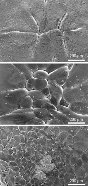 Figure 1: Scanning electron microscopy images showing the development of the chick embryo from eight open cells in yolk furrows (top) to laterally closed cell clusters (bottom). Reproduced from Ref. 1 and licensed under CC BY 3.0 © 2014 H. Nagai et al.
The mysterious period of early chick development that occurs inside the mother hen has been revealed for the first time by a collaborative team involving researchers from RIKEN, Seoul National University in Korea and Shizuoka University in Japan1. The study demonstrates that despite apparent differences, vertebrate embryos undertake very similar cellular journeys and share fundamentally conserved developmental mechanisms.
Early chick development was generally characterized in the 1970s, but no further cellular or molecular details of the crucial first 25 hours post-fertilization have been subsequently reported. As Guojun Sheng from the RIKEN Center for Developmental Biology explains: “The chick is notorious as the only model animal commonly used in developmental biology whose early phase of development is inaccessible for experimental analysis.”
Within 13 hours of fertilization, the fertilized chick egg develops through a rapid series of cell cycles to produce cell clusters. This is known as the ‘cleavage’ stage of embryonic development; molecular and cellular events taking place during this stage are crucial for later lineage specification. Little is known, however, about these events in chick embryos. For example, like other vertebrates that develop from a yolk-rich egg, chick embryos begin to direct their own development relatively early, but the exact timing of this transition, known as zygotic gene activation, is unknown.
To investigate, Sheng and his colleagues collected early cleavage stage chick embryos from laying hens using a non-invasive abdominal massage method and examined the embryos by scanning electron microscopy. The research team was able to reconstruct the development of the embryo over the course of a few hours, from 8 open cells nestled in furrows in the yolk surface, to 16 closed cells that then kept multiplying to form a four-cell-thick layer of cell clusters (Fig. 1). They also observed the formation of a cavity above the yolk surface and a yolk syncytial layer similar to that observed in fish embryos.
Using a marker of gene transcription, Sheng and his colleagues were able to determine that zygotic gene activation takes place after about eight cell cycles in chick embryos. In mammals, zygotic gene activation takes place before the third cell cycle, whereas in fish and frog embryos, it does not occur until the eighth or ninth cell cycle. The results suggest the existence of an evolutionary conserved mechanism for control of zygotic gene activation in yolk-rich embryos. “My aim is to convince people that the chick is a great model for understanding early human development,” says Sheng.
References
- 1. Nagai, H., Sezaki, M., Kakiguchi, K., Nakaya, Y., Lee, H. C., Ladher, R., Sasanami, T., Han, J. Y., Yonemura, S. & Sheng, G. Cellular analysis of cleavage-stage chick embryos reveals hidden conservation in vertebrate early development. Development 142, 1279–1286 (2015). doi: 10.1242/dev.118604