Oct. 30, 2015
Scrutinizing biological nanomachines
Saori Maki-Yonekura, Research Scientist
Please briefly describe your current research.
I study biological macromolecular structures, which include membrane proteins and macromolecular complexes. In bacteria, membrane protein complexes are involved in the active transport of essential nutrients, such as iron and vitamins, across the outer membrane. The mechanisms employed by these tiny nanomachines could be used to develop engineered nanosystems, but they are difficult to analyze. To tackle this problem, I combine various techniques such as x-ray and electron crystallography as well as cryo-electron microscopy (cryo-EM) to study the structures of biological macromolecules.
What has been the most interesting discovery in your field in the last few years?
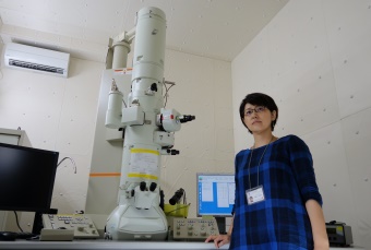
In cryo-EM, biological macromolecular structures are frozen in a thin layer of ice and imaged using an electron microscope. The three-dimensional structure of the macromolecular complex is then reconstructed by processing the images using a method called single-particle analysis. Cryo-EM is a very powerful technique because samples do not need to be crystallized, but its resolution has been limited until recently. By introducing high-resolution and fast-read-out electron detectors, which can correct for the movement of molecules in ice during data collection, researchers have been able to achieve resolutions comparable to, or even surpassing, those of x-ray crystallography.
Electron crystallography of thin three-dimensional (3D) crystals also provides a powerful means for determining the structures of crystals that are too small to observe using x-ray crystallography. This is because protein atoms diffract electrons four to five orders of magnitude more strongly than x-rays. The technique can also be used to visualize the charged states of amino acid residues and metals—information that cannot be obtained by x-ray crystallography.
 © 2015 RIKEN
These two new technologies—single-particle cryo-EM and electron 3D crystallography—could help to reveal the working mechanism of biological macromolecules in more detail. I am applying both methods to study the structures of membrane proteins under different physiological conditions.
How did you become interested in your current field of research?
After completing my master’s degree, I worked at a research institute that had the latest electron microscope. I dreamt of using this microscope to determine high-resolution structures of biological macromolecular complexes. Over the course of my research career, I have realized that the combined approach of x-ray crystallography and cryo-EM is very powerful for studying the structures of difficult biological targets.
More recently, I have started using the facilities at the RIKEN SPring-8 Center to combine x-rays and electron beams for structural biology. These include the high-brilliance x-ray sources produced by beamlines BL32XU and BL41XU at the SPring-8 synchrotron radiation facility, the SPring-8 Angstrom Compact free electron LAser (SACLA), and the automated web-based crystal screening system and data collection for x-ray diffraction data, called D-Cha.
How has being at RIKEN helped your research?
RIKEN offers a suitable environment for researchers to concentrate on their studies. Despite belonging to a small laboratory, I can learn about the latest techniques from the many experts in x-ray crystallography at the RIKEN SPring-8 Center.