Apr. 27, 2018 Research Highlight Biology
Structural insights into a protein-modification mechanism that boosts bacterial virulence
The molecular structure of a bacterial enzyme reveals how a sugar is added to a protein that supports the growth of pathogenic bacteria
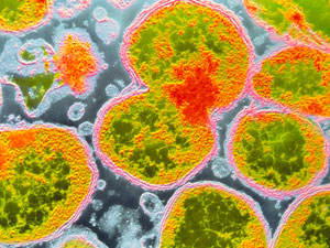 Figure 1: Insights into the structure and workings of a bacterial enzyme could lead to new antibiotics for the Neisseria meningitidis bacterium (shown in micrograph), which causes meningitis. CNRI/SCIENCE PHOTO LIBRARY
The molecular structure of an important enzyme in the disease-causing bacteria Neisseria meningitidis has been determined by a team of RIKEN researchers1. This information sheds light on how the enzyme works and also highlights targets for new antibiotics against several diseases, such as meningitis, gonorrhoea and pertussis.
The addition of sugar molecules to proteins regulates many cellular processes. Somewhat surprisingly, most enzymes that facilitate this addition (known as glycosyltransferases) can modify a variety of molecules. This is because their structures recognize the overall shape of protein domains and do not support the formation of many direct interactions with the protein’s conserved amino acid residues.
An earlier study had revealed how the bacterial glycosyltransferase EarP transferred the naturally occurring sugar rhamnose to Arginine 32 of translation elongation factor P (EF-P). This unusual protein modification mechanism activates EF-P, which spurs on ribosomes that have stalled before protein synthesis is complete and stimulates the production of proteins that are toxic to the infected host.
To further understand how EF-P is activated, a team led by Shigeyuki Yokoyama and Tatsuo Yanagisawa of the RIKEN Structural Biology Laboratory analyzed the structure of N. meningitidis EarP. They showed with unprecedented detail how EarP recognizes its substrate protein (EF-P) and changes its shape, thereby allowing the sugar transfer reaction to occur.
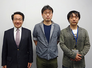 Shigeyuki Yokoyama (left), Tatsuo Yanagisawa (center) and Toru Sengoku (right), with co-workers, have revealed the structure and mechanism of action of arginine rhamnosyltransferase (EarP) in Neisseria meningitides. © 2018 RIKEN
Unlike other protein glycosyltransferases whose structures have been characterized, EarP binds to a particular region of EF-P, the β-sheet structure of EF-P domain I, through many interactions that recognize its conserved amino acid residues. In the absence of EF-P, EarP binds to a rhamnose donor in an inactive shape that does not allow the sugar to transfer to another protein. EF-P binding induces a shape change that allows the transfer of rhamnose to EF-P’s Arginine 32 and the activation of EF-P.
“This novel and sophisticated mechanism ensures that EarP’s only authentic substrate is rhamnosylated,” explains Yokoyama.
Interestingly, most of the amino acid residues in EarP involved in the binding with EF-P and the rhamnose donor are conserved among EarP-containing bacteria. Mutating these residues in EarP significantly reduced its sugar transfer activity, which suggests that they could be a good starting point for developing EarP inhibitors.
The researchers anticipate their findings will help the development of new antibiotics. “A detailed understanding of the EarP structure and its unique reaction mechanism enables the rational, structure-based design of first-in-class drug candidates specific to a group of pathogenic bacteria,” says Yokoyama.
Related contents
- Blocking obesity with a protein-sugar combination
- Mapping a metabolic master switch
- Unraveling a highly specific protein modification
References
- 1. Sengoku, T., Suzuki, T., Dohmae, N., Watanabe, C., Honma, T., Hikida, Y., Yamaguchi, Y., Takahashi, H., Yokoyama, S. & Yanagisawa, T. Structural basis of protein arginine rhamnosylation by glycosyltransferase EarP. Nature Chemical Biology 14, 368–374 (2018). doi: 10.1038/s41589-018-0002-y