Sep. 21, 2010 Press Release Biology Chemistry
World's largest oligosaccharide cluster reveals influence of molecular structure on metabolism
Experiments using a new type of synthetic cluster with a world record molecular weight of more than 50,000 have revealed the dramatic impact of oligosaccharide structure on the dynamics of metabolic uptake. Developed by researchers at the RIKEN Center for Molecular Imaging Science (CMIS), Osaka University, and Kishida Chemical Co. Ltd., the clusters open the door to new oligosaccharide-based imaging tracers for diagnosing diseases such as cancer.
Asparagine-linked oligosaccharides (N-glycans), carbohydrates that link to proteins via the amino acid asparagine (Asn), play an essential role in a wide variety of cellular functions including cell-cell recognition, cell adhesion and quality control. The complex branching structures that many N-glycans form on the cell surface facilitate such functions by increasing the likelihood of specific cell, protein and oligosaccharide interactions. Little is known, however, about what structural features enable N-glycan clusters to do this.
To investigate this question, the researchers designed a series of artificial polylysine-based N-glycan clusters, the largest made up of 16 N-glycans, by mimicking their natural bio-environment. Unlike earlier research on N-glycans conducted in artificial "in vitro" environments, the team studied the synthetic clusters in live mice, a natural setting offering unique insights into biological function.
Using positron emission tomography (PET) and in vivo fluorescence imaging, the team analyzed how glycoclusters labeled with the radioligand68Ga-DOTA and fluorophore Cy5 moved through various organs of the body. While small glycoclusters were quickly excreted, in the largest glycocluster, the presence of a specific sialic acid linkage to galactose, a type of sugar, altered the fates of clusters entirely: those with the linkage accumulated in the liver, while those without it were cleared through the kidney to the urinary bladder.
Reported in the German journal Angewandte Chemie International Edition, the findings clarify for the first time how particular structural features of N-glycans result in their accumulation in specific organs, paving the way for the development of new synthetic glycoclusters and more efficient tracers for molecular imaging.
Reference
- Katsunori Tanaka, Eric R. O. Siwu, Kaori Minami, Koki Hasegawa, Satoshi Nozaki, Yousuke Kanayama, Koichi Koyama, Weihsu C. Chen, James C. Paulson, Yasuyoshi Watanabe, and Koichi Fukase. Non-Invasive Imaging of Dendrimer-type N-Glycan Clusters: Remarkable In Vivo Dynamics Dependence on Oligosaccharide Structure. Angewandte Chemie International Edition (2010).
Contact
Yasuyoshi Watanabe
Satoshi Nozaki
Koki Hasegawa
Molecular Probe Dynamics Laboratory
RIKEN Center for Molecular Imaging Science
Tel: +81-(0)78-304-7124 / Fax: +81-(0)78-304-7126
Jens Wilkinson
RIKEN Global Relations and Research Coordination Office
Tel: +81-(0)48-462-1225 / Fax: +81-(0)48-463-3687
Email: pr@riken.jp
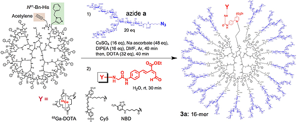
Figure 1: Preparation of N-glycan clusters through histidine accelerated Cu(I)-mediated Huisgen 1,3-dipolar cycloaddition and labeling by 6π-azaelectrocyclization
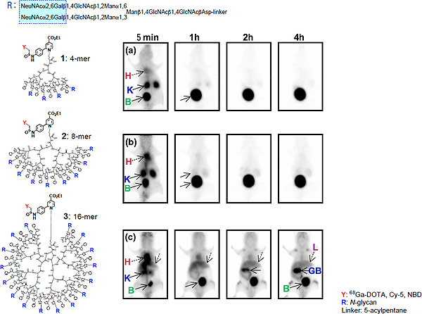
Figure 2: Dynamic PET imaging of glycoclusters in normal BALB/c nude mice.68Ga-DOTA-Labeled glycoclusters were administrated from the tail vein of the mice and the whole body was scanned by a small animal PET scanner at intervals over a 4 hour period after injection.
Top (a): tetra-glycocluster (4-mer) made up of 4 N-glycans
Middle (b): octa-glycocluster (8-mer) made up of 8 N-glycans
Bottom (c): hexadeca-glycoclusters (16-mer) made up of 16 N-glycans
H: heart; K: kidney; L: liver; B; urinary bladder; GB: gallbladder
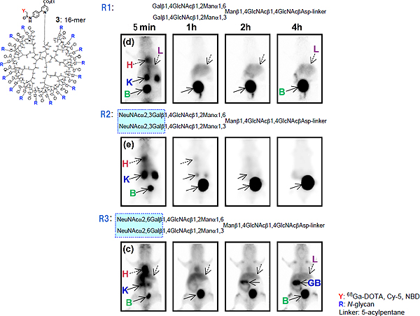
Figure 3: Dynamic PET imaging of 16-mer glycoclusters with different structural features
R1: asialo-glycan.
R2: Neuα(2-3)Gal-glycan.
R3: Neuα(2-6)Gal-containing glycan.
H: heart; K: kidney; L: liver; B; urinary bladder; GB: gallbladder.