Apr. 26, 2016 Press Release Biology
Change in the brain: astrocytes finally getting the recognition they deserve
Researchers at the RIKEN Brain Science Institute (BSI) in Japan have demonstrated that astrocytes help control the strength of connections between neurons. Published in Proceedings of the National Academy of Sciences, the study used cultured cells and brain slices to show that astrocytes in the hippocampus regulate changes in the brain brought on by neural activity.
Whenever we learn something new or are affected by our experiences, it’s because synapses — the connections between neurons in our brain — have changed. Sometimes new synapses are made, and other times the strengths of existing synapses are turned up or down. When synapses are strengthened, the signal from one neuron to the next results in a bigger response than it used to, and vice versa. Until recently, synaptic strength was thought to change only at synapses of active presynaptic neurons. Now, RIKEN scientists have shown that the truth is more complicated, and more interesting.
“We have found an active mechanism that helps to increase variation in synaptic strength,” explains lead scientist Yukiko Goda, “and surprisingly, it comes from astrocytes, which have previously been thought to play mostly passive roles in the brain.”
Astrocytes are a type of non-neural glial cell in the brain, often described as support cells for neurons. While recent studies have shown that they might have some global effects on neuronal transmission, Goda’s study shows that they can have very local effects at the individual synapse level.
To examine the effects of astrocyte activity on synaptic strength, the team first set up a culture of hippocampal neurons and astrocytes. Then they found two neurons that each connected to a third target neuron at a separate synapse — but were not connected with each other — and examined the strengths of the two synapses under different conditions.
Expected changes in synaptic strength were found at a synapse when the presynaptic neuron connected to it was stimulated with electrical pulses. At the same time, they found that this was often accompanied by changes at the other, non-stimulated, synapse.
Testing showed that the changes at these “heterosynapses” were not related to the postsynaptic neuron, but were blocked by an NMDA receptor antagonist. Further testing showed that blocking astrocyte activity also prevented changes at the heterosynapses.
But what happens when neurons are not being experimentally stimulated?
In this case, they found that blocking astrocyte NMDA receptors by any of several methods caused synaptic strengths of converging inputs on a given neuron to become more equal, whether in culture or in intact hippocampal slices. As Goda explains, “we found that astrocyte activity helps maintain normal variation of synaptic strengths, even when strong, plasticity inducing stimulation is absent.”
Understanding how astrocytes regulate synaptic strength, as well as the importance of this type of plasticity is not merely academically important.
“As synaptic dysfunction is thought to trigger or exacerbate many neurological diseases, a deeper understanding of how synaptic communication is regulated will aid in discovering disease mechanisms and developing treatments. Our work shows that astrocytes could be a potential target of novel therapeutics.”
“Our next goal,” continues Goda, “is to determine the precise signaling mechanism by which astrocytes target presynapses, and whether communication between astrocytes is also involved in controlling synapse variability.”
Reference
- Mathieu Letellier, Yun Kyung Park, Thomas E Chater, Peter H Chipman, Sunita Ghimire Gautam, Tomoko Oshima-Takago, Yukiko Goda, "Astrocytes regulate heterogeneity of presynaptic strengths in hippocampal networks", Proceedings of the National Academy of Science, doi: 10.1073/pnas.1523717113
Contact
Laboratory Head
Yukiko Goda
Laboratory for Synaptic Plasticity and Connectivity
RIKEN Brain Science Institute
Adam Phillips
RIKEN International Affairs Division
Tel: +81-(0)48-462-1225 / Fax: +81-(0)48-463-3687
Email: pr@riken.jp
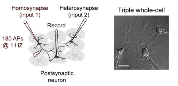
Experimental setup
(left) Schematic of the experimental setup in culture. (right) Differential interference contrast (DIC) image of 12-days-in-vitro cultured hippocampal neurons during a triple recording. Scale bar, 40 μm.
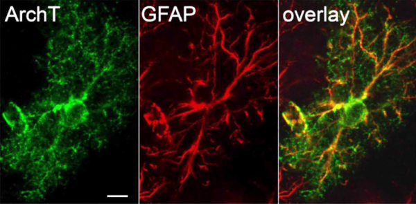
Astrocyte labeling for optogenetic inhibition
Astrocytespecific expression of ArchT-GFP (green) throughout the GFAP-positive processes (red) with stereotyped astrocyte morphology. Astrocytes were hyperpolarized optogenetically, causing variability of synapse strength to be reduced (as measured by the correlation in paired-pulse ratios at each synapse).