Jul. 5, 2019 Research Highlight Biology
Analyzing chromatin structure in more detail than ever before
The ability to explore the genome on a sub-nucleosome level promises to lead to new discoveries about how chromatin structure is linked to epigenetics
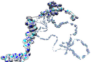 Figure 1: RIKEN researchers developed a technique that can reveal the positions and orientations of nucleosomes in the yeast genome at high resolution. © 2019 RIKEN Center for Biosystems Dynamics Research
A powerful method that can pinpoint the positions and orientations of individual nucleosomes—one of the building blocks of chromatin, which contains the genetic material of a cell—has been demonstrated on yeast by a RIKEN team1. It will help researchers to explore the relationship between chromatin structure and function.
While the molecular structure of DNA has been known for over 60 years, more recent research has shown how DNA is packaged into cell nuclei as thread-like chromatin interspersed with nucleosome ‘beads’. But despite recent advances, there is still a blind spot in our understanding of genome structure, namely the details of how nucleosomes are folded. Such knowledge is important because nucleosome positioning and orientation can couple with molecular reactions in the genome that cause gene regulation and other genome functions.
An analytic technique known as high-throughput chromosome conformation capture (Hi-C) can reveal information about large-scale chromatin structure, but it cannot show structure on the sub-nucleosome level.
“The three-dimensional nucleosome folding structure across the genome was still uncovered,” comments Yuichi Taniguchi of the RIKEN Center for Biosystems Dynamics Research (BDR). “If we can achieve a nucleosome-level resolution, I anticipate we will see a very strong connection between genome structure and function. I think it’s the next frontier in genetics.”
 Yuichi Taniguchi (middle of back row) and his team have used high-throughput chromosome conformation capture and molecular dynamics simulations to analyze the 3D genome-wide nucleosomal structure of yeast chromatin. © 2019 RIKEN
The low resolution of Hi-C means it cannot reveal the orientations of nucleosomes—an important aspect of chromatin structure. “Just like you can’t play dominoes without specifying the orientation of each domino tile, you can’t know chromatin structure without knowing the nucleosome orientations,” says David Priest, who was a postdoctoral researcher at BDR at the time of the study.
Now, by combining Hi-C with molecular dynamics simulations, Taniguchi and colleagues have been able to reveal the positions and orientations of nucleosomes in the yeast genome at high resolution (Fig. 1).
“The crucial advance in our method over previous Hi-C methods is that it can measure interactions not just between whole nucleosomes, but also between the DNA entry and exit points of the nucleosome,” explains Priest. “Since the DNA enters and exits on approximately opposite sides of the nucleosome, this sub-nucleosome interaction data will allow us to map nucleosome orientations.”
Using their technique, the researchers discovered that the nucleosomes in yeast exhibit two folding motifs: four adjacent nucleosomes can form a pyramid-like structure or a flat rhombus. This finding could explain two competing models of chromatin structure proposed in earlier studies.
The team intends to apply the technique to more complex situations. “Our next challenge is to apply the technique to the human genome and expand our findings to more general cases,” says Masae Ohno, the first author of the study.
Related contents
- Elongation factors smooth transcription in the nucleosome
- Listening to the noise
- Molecular structure of elongation complex essential for DNA transcription determined
Video
References
- 1. Ohno, M., Ando, T., Priest, D. G., Kumar, V., Yoshida, Y. & Taniguchi, Y. Sub-nucleosomal genome structure reveals distinct nucleosome folding motifs. Cell 176, 520–534 (2019). doi: 10.1016/j.cell.2018.12.014