Feb. 16, 2007 Research Highlight Biology
Not just counting sheep
Research has revealed neuronal firing patterns that could help explain how the brain makes memories during deep sleep
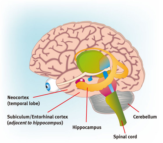 Figure 1: Regions of the brain.
Most people think of a good night’s rest as a refreshing and relaxing experience, but even with closed eyes and a still body, the brain continues to work away. In fact, many neuroscientists believe that the brain uses certain stages of sleep—so-called ‘slow-wave’ sleep—as an opportunity to process the day’s events.
A key component of this process is communication between the hippocampus, the brain’s primary memory center, and the neocortex, which handles a wide range of brain functions, including sensory processing. “Memory information may be consolidated in the hippocampus and transferred to the neocortex during sleep, especially slow-wave sleep,” explains Yoshikazu Isomura of the RIKEN Brain Science Institute in Wako. “In fact, neuronal firing patterns experienced in a waking and behaving state are often reproduced during slow-wave sleep.”
Previous research has shown that neocortical neurons exhibit slow oscillatory patterns of activity during slow-wave sleep, between a highly active 'UP' state, and a hyperpolarized quiescent 'DOWN' state. However, it has been unclear exactly how this stage affects the behavior of neurons in the hippocampus and adjacent parahippocampal regions. To investigate this further, Isomura and colleagues at Rutgers University in New Jersey performed a series of experiments to characterize the firing patterns of single neurons and groups of neurons in anesthetized and naturally sleeping rats1.
They uncovered evidence that although parahippocampal neurons, such as those in the entorhinal cortex and subiculum (Fig. 1), undergo slow oscillations that synchronize with those occurring in the neocortex, most hippocampal neurons do not. Nevertheless, hippocampal neurons were influenced by the activity of entorhinal neurons in the ‘UP’ state, and even exhibited subarea-specific activity when entorhinal and neocortical neurons were in the ‘DOWN’ state. Isomura and his colleagues postulate that this communication from neocortex to hippocampus may trigger the ‘sharp-wave-ripple’ hippocampal oscillations that signal back to the neocortex.
“Researchers in this field believe that sharp-wave-related ripple oscillations may contribute to memory consolidation in the hippocampus or memory transfer from the hippocampus to the neocortex,” Isomura says.
These findings could provide an important piece in the puzzle of understanding memory processing during deep sleep, but there are still many unanswered questions. In particular, although the entorhinal cortex appears to play a role in this memory-building process, the specifics remain unclear. “The entorhinal cortex is not so well understood, even in its basic physiological properties,” Isomura says, “and we will focus on its functional role in relation to hippocampus–neocortex interaction in the near future.”
References
- 1. Isomura, Y., Sirota, A., Özen, S., Montgomery, S., Mizuseki, K., Henze, D.A. & Buzsáki, G. Integration and segregation of activity in entorhinal-hippocampal subregions by neocortical slow oscillations. Neuron 52, 871–882 (2006). doi: 10.1016/j.neuron.2006.10.023