Oct. 24, 2007 Research Highlight Biology
How eating cell ‘corpses’ reduces inflammation
Specialized immune cells orchestrate proper elimination of dead cells to prevent inflammation
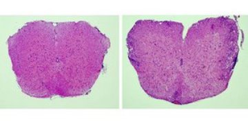 Figure 1: Normal (left) and inflamed spinal cord (right). The multiple sclerosis-like disease occurs when specialized macrophages that prevent inflammation are depleted. © J. Clin. Invest./American Society for Clinical Investigation/117/2273 (2007)
Reporting in the August issue of The Journal of Clinical Investigation 1, a team of Japanese researchers has found that immune cells called ‘marginal zone macrophages’ prevent inflammation by promoting the elimination of cells that have just died—so-called cell ‘corpses’.
It has been long known that certain types of dead cells can suppress inflammation. A cell ‘programmed’ to die goes through a tranquil process called apoptosis, whereas traumatically killed cells die by a process called necrosis. Only apoptotic corpses can suppress inflammation.
Led by Masato Tanaka at the RIKEN Research Center for Allergy and Immunology, Yokohama, the team observed that apoptotic corpses injected into experimental mice migrate to specific locations in the spleen and lymph nodes and then disappear—phenomena associated with suppression of experimentally-induced inflammation. Intriguingly, marginal zone macrophages are found in the same locations.
Testing whether the macrophages were important for the disappearance of the corpses and reduced inflammation, the team depleted the macrophages from mice and then injected apoptotic cells. They found that the corpses were present much longer and experimentally-induced brain inflammation could no longer be suppressed (Fig. 1).
Digging deeper to understand this, the team looked at other nearby immune cells and found differences in two types of cells called dendritic cells, one of which was known to suppress inflammation.
Studying how the two types of dendritic cells responded to apoptotic corpses when the macrophages were present or absent, Tanaka’s team found that the dendritic cell type known to suppress inflammation could do so only when the macrophages were present. In the absence of the macrophages, the other dendritic cells caused inflammation.
Further observations indicated a difference in the way the dendritic cells responded to the apoptotic corpses––which are normally ‘eaten’ by the inflammation-suppressing dendritic cells. The team noticed that when the macrophages were absent, the second type of dendritic cells could ingest the apoptotic corpses, which caused inflammation.
“We are now currently investigating the differences between the two [types of] dendritic cells,” says Tanaka. One possibility is that the specialized macrophages transport apoptotic corpses selectively to the dendritic cells that suppress inflammation, thus physically preventing the other type of dendritic cells from promoting inflammation.
Exactly how marginal zone macrophages and two types of dendritic cells effect this complex processing of apoptotic corpses remains unknown, says Tanaka. Nevertheless, the observations are clear and represent a potential interesting avenue of research in causes of inflammation.
References
- 1. Miyake, Y., Asano, K., Kaise, H., Uemura, M., Nakayama, M. & Tanaka, M. Critical role of macrophages in the marginal zone in the suppression of immune responses to apoptotic cell-associated antigens. The Journal of Clinical Investigation 117, 2268–2278 (2007). doi: 10.1172/JCI31990