Mar. 7, 2008 Research Highlight Biology
Brain repairs
Healing from spinal cord injury requires use of different regions of the brain
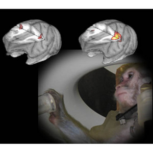 Figure 1: A test monkey in a PET scanner and two representative images during the early and late phases of recovery. After a spinal cord injury affecting the dexterous finger movement of the right forepaw, the early phase of retraining results in both the ipsolateral and contralateral sides of the brain being active (left image). During the later phase of recovery, the contralateral side of the brain is again the predominately active region when the right forepaw is in use, whereas activity in the ipsolateral brain regions has ceased (right image).
New research from Japan using non-human primates and a brain imaging technique called Positron Emission Tomography (PET) has shown that compensatory mechanisms in the brain contribute to recovery from spinal cord injury.
After partial injury to the spinal cord or brain, retraining to complete physical tasks, or neurorehabilitation, often relies on recruiting remaining intact brain regions to compensate for the injured neurons. But exactly which regions the brain uses to rewire itself, and when, has puzzled scientists.
Now, Hirotaka Onoe at the RIKEN Molecular Imaging Program of the Kobe Institute, in collaboration with Tadashi Isa at the Japanese National Institute for Physiological Sciences and colleagues, has used PET scanning to follow the recovery of injured monkeys to address this question1.
The researchers transected a neural pathway of three monkeys such that they could no longer use the fine motor skills of their right forepaws to carefully grasp a small piece of food. Finger dexterity, however, could recover with rehabilitation within three months.
PET scans showed that pre-injury, as occurs normally, only the opposite—or contralateral—side of the brain to the right forepaw is active when the limb is in use. However, after injury, the same—or ipsolateral—side of the brain as the right forepaw also becomes active to help compensate for the injured neuronal pathway.
Eventually, though, the ipsolateral activity declines as the injured neuronal pathway heals and the contralateral brain regions again become the predominant areas of activity (Fig. 1). Indeed, this compensation by the ipsolateral brain region is only needed in the initial stages of recovery as early inactivation of these ipsolateral regions impedes recovery, whereas inactivation at later times results in no impediment.
From these results the team has concluded that normally the activity of the ipsolateral side of the brain is inhibited when an appendage is in use, but after injury this inhibition is relaxed in order to allow some use of that limb and simultaneously allow the injured contralateral neuronal pathway to recover. However, once sufficient recovery of the injured neuronal pathway has occurred—either by restoration of the original wiring or recruitment of other nearby wiring as a workaround solution—the inhibition of the ipsolateral brain region returns.
Onoe and colleagues hope that by better understanding this recovery process it will be possible to use PET scanning to follow the progress of spinal cord injury patients during their rehabilitation and predict which ones might be good candidates for recovery.
References
- 1. Nishimura, Y., Onoe, H., Morichika, Y., Perfiliev, S., Tsukada, H. & Isa, T. Time-dependent central compensatory mechanisms of finger dexterity after spinal cord injury. Science 318, 1150–1155 (2007). doi: 10.1126/science.1147243