May 16, 2008 Research Highlight Biology
How nerve cells are shaped
Discovery of molecules that sculpt nerve shape will assist in understanding nerve cell function and neurological disease
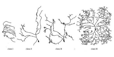
 Figure 1: Shapes of the four classes of sensory nerve cells in Drosophila. Reproduced with permission from Ref. 1 © 2007 Elsevier Inc.
Molecular biologists at RIKEN’s Brain Science Institute in Wako have unraveled details of the genetic controls that determine the distinctive shapes of four classes of sensory nerve cells in the fruit fly, Drosophila.
Nerve cell shapes vary according to the number, branching and disposition of their projections or dendrites, collectively known as arborization. This determines their capacity for interacting with their environment and with other nerve cells or neurons, hence their computational ability and roles. Knowing how such shapes are determined is important for understanding nerve cell function and neurological disease.
The shapes of Drosophila sensory cells display a progression of increasing branch complexity and symmetry from class I to class IV (Fig. 1). The research, recently published in Neuron 1, shows how development of these shapes depends on the levels and interaction of transcription factors, which are proteins that control the information printed off from the DNA.
From previous work by other researchers it was known that only the transcription factor ‘Abrupt’—which suppresses outgrowth and branching of dendrites—is active in class I neurons. In the other three neuron classes Abrupt is switched off and the factor ‘Cut’ is active. Cut promotes growth and branching and is found at low levels in class II neurons, high in class III and intermediate in class IV.
Because class IV neurons have lower levels of Cut than class III, but are much larger and more highly branched, the researchers hypothesized the involvement of a third transcription factor in this biggest neuron class. They subsequently found ‘Knot’, which encourages dendrite outgrowth and branching primarily through promoting the protein Spastin. By transferring Knot and Cut into cells where they are not normally active, the group found that both regulate development of the cell skeleton, but each controls a different skeletal building block.
Knot and Cut interact in complex ways. In class III neurons, which lack Knot, spikes known as filopodia project from the main dendrite trunks. In class IV neurons where both factors are present, filopodia formation is suppressed by Knot. In contrast, the two factors work together to increase outgrowth and branching.
“We have begun to unravel the complex interactions of the many proteins controlling the characteristic shape of different neuron types,” says project leader, Adrian Moore. “But we have a long way to go to understand exactly how these molecular mechanisms translate into the final form of the neuron.”
References
- 1. Jinushi-Nakao, S., Arvind, R., Amikura, R., Kinameri, E., Liu, A.W. & Moore, A.W. Knot/Collier and Cut control different aspects of dendrite cytoskeleton and synergize to define final arbor shape. Neuron 56, 963–978 (2007). doi: 10.1016/j.neuron.2007.10.031
