Jan. 22, 2010 Research Highlight Biology
Switching neurons
Inhibitory neurons in the visual cortex of the brain exhibit a bidirectional form of plasticity after visual deprivation
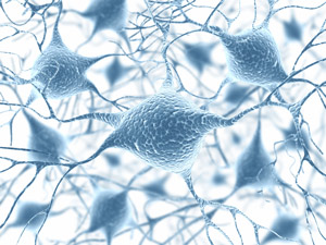
 Figure 1: Artistic representation of inhibitory interneurons in the visual cortex of mammals, which affect the response of pyramidal neurons to visual stimuli. © (2009) istockphoto/ ktsimage
If both eyes are open during mammalian development, pyramidal neurons in the visual cortex mature such that they ‘preferentially’ fire in response to the visual stimuli of one particular eye. If that eye is occluded during a critical period of development, the pyramidal neurons switch and fire in response to visual stimuli of the non-occluded eye. This leads to a loss of representation of the occluded eye in the visual cortex and the loss of visual acuity in that eye.
Pyramidal neurons receive inputs from so-called inhibitory interneurons within the visual cortex (Fig. 1), but it has been unclear how the interneurons respond to visual deprivation, and what their role is in inducing this plasticity of pyramidal neuron response. Now, a team led by Takao Hensch at the RIKEN Brain Science Institute (BSI) in Wako, has reported that inhibitory neurons also change their responsiveness to visual stimuli after one eye is occluded1. Surprisingly, unlike pyramidal neurons, which have a unidirectional change in responsiveness—only towards the open eye—the inhibitory neurons have a bidirectional change: initially, they respond preferentially to stimuli presented to the occluded eye, but later, they switch their responsiveness to the open eye.
In mice in which both eyes were open, Hensch and colleagues found that blocking the signals going from the inhibitory neurons to the pyramidal neurons caused the pyramidal neurons to lose their selective responsiveness to one eye. When they occluded one eye, blocking the inhibitory neuron signals caused the pyramidal neurons to flip their responsiveness from one eye to the other. This indicates that the signals sent from the inhibitory neurons to the pyramidal neurons help to control the response of pyramidal neurons to both normal vision and visual deprivation.
After the researchers determined how the neurons would react to changes in visual stimulation, they developed a network model to understand how connectivity between neurons—and the plasticity of these connections—would explain the responses of the pyramidal neurons and the inhibitory interneurons. The model, developed in collaboration with Tomoki Fukai and team also at BSI, demonstrates how individual components of a neuronal cell circuit contribute to plasticity within the brain.
“We will now pursue the detailed mechanisms of this plasticity to determine sites for therapeutic interventions,” says Hensch. Understanding how this cell type determines early brain plasticity offers the potential for cell-specific strategies to restore or reactivate proper brain function in several neurological disorders such as autism and schizophrenia.
References
- 1. Yazaki-Sugiyama, Y., Kang, S., Cateau, H., Fukai, T. Hensch, T.K. Bidirectional plasticity in fast-spiking GABA circuits by visual experience. Nature 462, 218–221 (2009). doi: 10.1038/nature08485
