Mar. 26, 2010 Research Highlight Biology
Eyes wide shut
Different cell types in the visual cortex respond differently to changes in visual experience
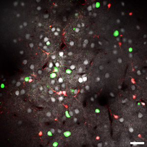 Figure 1: A two-photon calcium image of the visual cortex of a mouse, at a depth of 120 µm from the cortical surface. Inhibitory interneurons are shown in green and excitatory neurons are shown in white; red cells are astrocytes (scale bar, 30 µm). Reproduced from Ref. 1 © 2010 the authors
In the primary visual cortex of the brain, neurons are organized into alternating columns that receive inputs from either the left or right eye. This organization is strongly dependent on early visual experience. When one eye is deprived of visual inputs during a critical developmental period, the corresponding columns fail to develop properly, whereas those receiving inputs from the unaffected eye grow larger than normal.
The cortex consists primarily of two different types of neuron: excitatory neurons that synthesize and release the neurotransmitter glutamate, and inhibitory interneurons which use the transmitter γ-aminobutyric acid (GABA). How each of these cell types contributes to experience-dependent changes in the visual cortex is, however, unknown.
To investigate this, Tadaharu Tsumoto of the RIKEN Brain Science Institute, Wako, and his colleagues injected a calcium-sensitive dye, Fura-2, into the visual cortex of genetically engineered mice whose inhibitory interneurons express a fluorescent protein called Venus1. The intensity of Fura-2 fluorescence changes in response to the increase in calcium ion concentration that is characteristic of neuronal activity.
This approach enabled the researchers to both identify the interneurons in the visual cortex and monitor their activity (Fig. 1). In animals reared normally, they first identified the ‘binocular’ regions of the primary visual cortex by visually stimulating each eye in turn, and using two-photon laser-scanning microscopy to locate the cells that responded to both. This revealed that inhibitory interneurons are more responsive to inputs from both eyes than excitatory neurons.
The responses in mice deprived of visual inputs to one eye for two days during the critical period were then examined. The change in the responses of both cell types was found to be similar—both had become more responsive to inputs from the open eye.
When mice were deprived of visual inputs to one eye after the critical period, however, the effect observed was far stronger on the inhibitory interneurons. They tended to receive inputs from the open eye, and their responses to inputs from the closed eye were also depressed, whereas those of the excitatory neurons remained almost stable. The interneurons normally act to inhibit the excitatory neurons, so their depressed responses may contribute to the stability of excitatory neuron responses to the deprived eye.
“Inhibitory interneurons are divided into several subtypes,” says Tsumoto. So, the next step is to determine which particular subtypes are involved in maintaining plasticity after the critical period.
References
- 1. Kameyama, K., Sohya, K., Ebina, T., Fukuda, A., Yanagawa, Y. & Tsumoto, T. Difference in binocularity and ocular dominance plasticity between GABAergic and excitatory cortical neurons. Journal of Neuroscience 30, 1551–1559 (2010). doi: 10.1523/JNEUROSCI.5025-09.2010