Mar. 30, 2012 Research Highlight Biology
Measuring attention to detail
Human attention to a particular portion of an image alters the way the brain processes visual cortex responses to that image
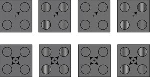 Figure 1: A schematic diagram of the contrast discrimination task, showing the focal cue trial (top row) and the distributed cue trial (bottom row). The contrast within the top right circle increases from the first interval (second column) to the second interval (fourth column). The third column is the interstimulus interval. Reproduced, with permission, from Ref. 1 © 2011 Elsevier Inc.
Our ability to ignore some, but not other stimuli, allows us to focus our attention and improve our performance on a specific task. The ability to respond to visual stimuli during a visual task hinges on altered brain processing of responses within the visual cortex at the back of the brain, where visual information is first received from the eyes. How this occurs was recently demonstrated by an international team of researchers led by Justin Gardner at the RIKEN Brain Science Institute in Wako1.
In a contrast discrimination task, the researchers showed three observers a stimulus of a group of four circles, each containing grey and white bars that created stripes of different contrasts (Fig. 1). After a short pause, the researchers showed the circles again, but the contrast within one of the circles was different. The observers were instructed to choose which group of circles contained the higher contrast.
In ‘focal cue trials’, an arrow directed the observers’ attention to a particular circle. In ‘distributed cue trials’, four arrows directed their attention diffusely, across all four circles. Gardner and colleagues found that the observers’ performance was better in the focal cue trials.
Using a magnetic resonance imaging (MRI) scanner, the research team was able to map the precise location within the visual cortex that was activated by the visual information within each of the four circles. During the contrast discrimination task, Gardner and colleagues could therefore measure the observers’ visual cortex activity elicited by the stimuli. In this way, they could correlate brain activity in the visual cortex with the observers’ attention and their choice of contrasting circles.
Visual cortex responses tended to be largest when the observers were paying attention to a particular target circle, and smallest when they were ignoring a circle. The researchers determined that the largest visual cortex responses to the stimuli guided the eventual choice of each observer, leading to enhanced performance on the visual task.
“We used computational modeling to test various hypotheses about how attention affects brain processing of visual information to improve behavioral performance,” explains Gardner. “We concluded that the observers’ attention causes their brains to select the largest cortical response to guide contrast choice, since we found that an ‘efficient selection’ model best explained the behavioral and fMRI data,” he says.
If the findings extend to other senses, such as hearing, researchers may begin to understand how humans make sense of a perceptually cluttered world.
References
- 1. Pestilli, F., Carrasco, M., Heeger, D.J. & Gardner, J.L. Attentional enhancement via selection and pooling of early sensory responses in human visual cortex. Neuron 72, 832–846 (2011). doi: 10.1016/j.neuron.2011.09.025