Feb. 21, 2014 Research Highlight Physics / Astronomy
Twin x-ray pulses light up matter
The SACLA free electron laser can now deliver pairs of high-intensity x-ray pulses with different wavelengths
X-ray beams are invaluable tools for scientific research. The patterns they create after diffracting through a crystal can reveal the atomic structure of the material, and blasting a sample with pulses of x-rays can help to identify the sample’s composition or capture snapshots of the atomic processes underlying incredibly fast chemical reactions.
Toru Hara and colleagues from the RIKEN SPring-8 Center and the Japan Synchrotron Radiation Research Institute have now developed a technique that can produce pairs of very short x-ray pulses in which each pulse has a different wavelength1. The system promises to open up a new frontier in x-ray science, allowing researchers to probe the atomic world in unprecedented detail.
The x-ray pulses are produced at SACLA (SPring-8 Angstrom Compact Free Electron Laser), which forms part of the SPring-8 (Super Photon Ring-8 GeV) synchrotron radiation facility in Harima. The SACLA x-ray free electron laser (XFEL) accelerates a stream of electrons to close to the speed of light and passes it through a series of five-meter-long undulator channels containing alternating magnetic fields. This path causes the electrons to ‘wiggle’ from side to side, forcing them to eject some of their energy by emitting x-rays (Fig. 1). These tightly bunched x-rays are all in phase, just like a coherent laser beam, and normally have the same wavelength, which is determined by the size of the gap between the undulator magnets. The energy of the x-ray pulses ranges from 5 to 15 kiloelectronvolts, and the pulses are typically less than 10 femtoseconds in length. The SACLA XFEL is one of only two facilities in the world where researchers can use such powerful, ultrashort x-ray pulses to study matter.
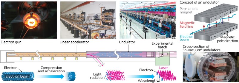 Figure 1: When bunches of electrons moving close to the speed of light are forced to wiggle through undulators, they produce coherent x-ray pulses.© 2014 RIKEN
Generating a double pulse
The new system generates twin pulses of different wavelengths by introducing a detour section between the eighth and ninth undulators. This magnetic chicane does not affect the x-rays generated in the first eight undulators, which continue on their course unimpeded. However, it forces the electrons to take a longer route, slightly delaying their arrival at the ninth and subsequent undulators. This second leg has a different magnetic gap, causing the production of x-rays of a different wavelength. The second x-ray pulse trails the first by up to 40 femtoseconds, and the delay can be fine-tuned to a fraction of a femtosecond. The resultant paired x-ray pulse train is referred to as a two-color double-pulse (TCDP) XFEL.
The intensity of each pulse can be adjusted by changing the number of operational undulators, and the two pulses can be made to hit a sample from slightly different directions by repositioning the second set of undulators. These features should make the x-rays useful for answering many different types of scientific questions, says Hara.
A new light source with many uses
 Figure 2: SACLA (SPring-8 Angstrom Compact Free Electron Laser) can generate intense pulses of x-rays to probe the atomic structure of matter. © 2014 RIKEN
The TCDP system has many potential applications in research. In imaging experiments, for example, the pulses could be used to produce complementary diffraction patterns that help to refine crystal structures with greater accuracy. “Taking images of a three-dimensional structure from different angles provides more information,” explains Hara.
When low-intensity x-rays are used, samples can be rotated and images taken from several angles. “In the case of XFELs, however, the sample is destroyed by just one intense pulse in most instances. So, many identical samples are prepared and exchanged for each XFEL shot,” notes Hara. Being able to take two snapshots at once would ease the burden of sample preparation.
The TCDP system could also be used to capture extremely fast events, such as structural changes in nanoparticles. Each x-ray pulse would produce a snapshot of the structure, and reveal how it evolves over a femtosecond timescale. This sort of technique could uncover the physical properties of materials at the atomic scale or track the course of chemical changes—vital information for researchers designing new materials and catalysts or trying to understand biological processes such as protein folding.
The two pulses could also serve different functions. The first could excite electrons in the target, and the second could record the outcome of that process immediately after using a ‘pump–probe’ approach. “In this case, the two pulses would need to have different wavelengths,” says Hara. “If the two pulses had the same wavelength, we would not be able to distinguish whether the observed signal was due to the pump pulse or the probe pulse.”
Greater separation makes the difference
Although other groups of researchers have also recently produced TCDP x-rays, the wavelengths of their two pulses only differ by a tiny percentage. “A separation of a few percent is not enough to clearly separate the signals,” says Hara. “Also, it limits the range of target processes that can be observed, since some processes need a large gap between the two wavelengths.”
In comparison, the wavelengths of the two x-ray pulses at the SACLA facility (Fig. 2) can be separated by more than 30 per cent. The wavelengths are also much shorter than achieved by other TCDP systems, providing better spatial resolution and revealing finer atomic details. The pulses have enough energy to rip electrons from the inner shells of atoms, triggering a cascade of other electrons that emit more x-rays in a characteristic spectrum that can be used to identify the atom—a technique known as energy-dispersive x-ray spectroscopy).
Researchers are now putting the system through its paces. “The two-color XFEL has already been used in experiments, but these are ongoing and the results have not yet been published,” says Hara. “I hope new ideas for experimental methods will emerge from these experiments in the future.”
References
- 1. Hara, T., Inubushi, Y., Katayama, T., Sato, T., Tanaka, H., Tanaka, T., Togashi, T., Togawa, K., Tono, K., Yabashi, M. & Ishikawa, T. Two-colour hard x-ray free-electron laser with wide tunability. Nature Communications 4, 2919 (2013). doi: 10.1038/ncomms3919
About the Researcher
Toru Hara

Toru Hara was born in Tokyo, Japan, in 1966. He graduated from the Faculty of Engineering at the University of Tokyo in 1989 and received his PhD from the University of Paris XI in France in 1995. He entered RIKEN as a research scientist for the JAERI–RIKEN SPring-8 Project Team, later joining the RIKEN SPring-8 Center. At the center, Hara contributed to SACLA (SPring-8 Angstrom Compact Free Electron Laser) and its precursor, the SPring-8 Compact SASE Source (SCSS), developing insertion devices and accelerator optics design as well as contributing to the operation of the free electron lasers. In 2011, Hara became team leader of the Beam Dynamics Team at the RIKEN SPring-8 Center. His current research focuses on the beam dynamics of linear accelerators and free-electron-laser physics.