Sep. 9, 2016
Clearing the brain
Meng-Tsen Ke, Foreign Postdoctoral Researcher

Laboratory for Sensory Circuit Formation, RIKEN Center for Developmental Biology
Please describe your current research.
I work on the application and assessment of optical clearing methods for imaging large brain tissues. Optical clearing enhances our ability to see deep into the brain by rendering tissue transparent. Combining these techniques with genetic labeling and advanced microscopy allows for the three-dimensional (3D) reconstruction of intact tissues such as neuronal circuits or solid tumor cell clusters.
How did you become interested in this field of research?
I started my PhD by dissecting neuronal circuits in the mouse olfactory system responsible for the sense of smell. But the lack of a suitable tissue clearing method made dissections difficult and time consuming. To reconstruct the mouse brain, I had to first slice samples into thin sections, image each section separately, and then carefully stitch them back together to get a total sum of the volume. The stitching process required a lot of corrections and modifications. To improve and speed up the flow of my experiments, I started to develop an optical clearing technique. This is just another example of a technology being invented out of necessity
What excites you the most about your research?
The boom in optical clearing methods has been impressive. Currently, optical clearing techniques enable the 3D imaging of intact tissue in fixed samples at cellular to synaptic resolutions. But new concepts and ideas are continually pushing the field forward. In the near future, in vivo optical clearing for in-depth imaging will enable us to investigate cellular activity or subcellular dynamics at deeper brain regions under physiological conditions.
What has been the most interesting discovery in your field in the last few years?
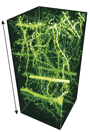 Optical clearing methods have enabled large-scale, super-resolution imaging of neural circuits in the mouse brain. © 2016 RIKEN
Recent developments in super-resolution microscopy have enabled us to inspect fine structural details in neuronal circuits at nanoscale spatial resolutions. This technology fills the gap between picometer-scale electron microscopy and micrometer-scale light microscopy for developing comprehensive maps of synaptic connections. As the availability of commercial super-resolution microscopy widens and optical clearing techniques improve, we should easily be able to obtain high-resolution 3D images of fluorescence-labeled samples, while saving time and effort on image alignment.
How and when did you join RIKEN?
I joined RIKEN in 2011 as a PhD student through the Joint Graduate School Program. After graduating from Kyoto University, I continued my research at the same laboratory through the Foreign Postdoctoral Researcher Program.
What is the best thing about working at RIKEN?
RIKEN is supportive and constantly encourages young scientists to pursue interesting, but risky, research topics. I am also impressed by the funding system and open atmosphere that is conducive to active discussions and interactions among researchers. RIKEN has given me access to the latest facilities and devices in the field.
Is there anything you wish you had known before coming to Japan?
As a Taiwanese, my transition to Japan was relatively smooth. Taiwan and Japan share cultural similarity compared with Western countries, and the common kanji characters have been useful in familiarizing me with the new language.