Sep. 6, 2011 Press Release Biology
New technique elucidates dynamics of plant cell metabolites
A new technique developed by researchers at RIKEN has clarified the location and dynamics of specific metabolites in a single cell of the alga Chara australis. The findings reveal that these metabolites are regulated and fluctuate under stress conditions, providing insight into previously-unknown functions of the vacuole in cellular processes.
Metabolites, the intermediates and products of chemical reactions that sustain all living organisms, play a central role in cellular processes including growth, differentiation and defense. Despite their importance, however, our understanding of the role of these metabolites in the cell is incomplete due to the lack of techniques for analyzing them at high spatial resolution.
In a paper to appear in the journal Plant Physiology, researchers at the RIKEN Plant Science Center (PSC) in Yokohama, Japan describe a novel technique which enables such high-resolution metabolite analysis. Their research focuses on Chara australis, an alga whose single, extremely large (up to 20 cm long) internodal cell provides a unique opportunity to study the detailed location and dynamics of metabolites inside the cell.
Their technique separates the cell into two parts, isolating its cytoplasm (containing plastids, mitochondria, nuclei, endomembrane system and cell wall) from its vacuole, a large water-filled compartment containing organic and inorganic molecules. Using this technique, the researchers demonstrate that the concentrations of 125 known metabolites in the vacuole and cytoplasm fluctuate asynchronously under stress conditions, suggesting that metabolites are spatially regulated in the cell.
By shedding light on the detailed distribution of metabolites in the cell, this finding marks a major advance in our understanding of plant cell metabolism. The technique used likewise charts new ground, providing unprecedented detail on organelle-specific metabolite concentration and highlighting the usefulness of C. autralis as a model organism for biological studies at the single-cell level.
Reference
Akira Oikawa, Fumio Matsuda, Munehiro Kikuyama, Tetsuro Mimura and Kazuki Saito, "Metabolomics of a single vacuole reveals metabolic dynamism in an alga Chara australis." Plant Physiology, 2011, DOI: 10.1104/pp.111.183772
Contact
Akira Oikawa
Metabolomic Function Research Group (Tsuruoka)
RIKEN Plant Science Center
Tel: +81-(0)235-25-3580 / Fax: +81-(0)235-25-3580
Kazuki Saito
Metabolomic Function Research Group
RIKEN Plant Science Center
Tel: +81-(0)45-503-9488 / Fax: +81-(0)45-503-9489
Jens Wilkinson
RIKEN Global Relations and Research Coordination Office
Tel: +81-(0)48-462-1225 / Fax: +81-(0)48-463-3687
Email: pr[at]riken.jp
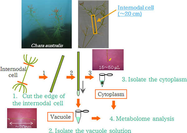
Figure 1: Isolation of vacuolar and cytoplasmic fraction from an internodal cell of C. australis.
The internodal cell (shown in the orange square in the upper-right photograph) is a large single cell growing to over 20 cm length. The vacuole solution of this cell is easily isolated by cutting the edge of the cell and emptying out its contents. The remaining cytoplasmic fraction and the vacuole solution are used for metabolomic analysis.
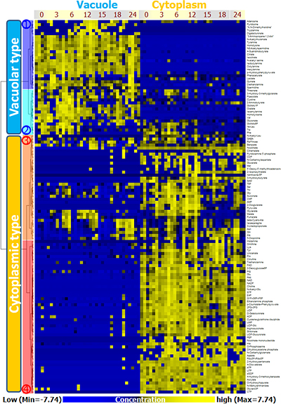
Figure 2: Localization of metabolites in a cell and fluctuation of metabolite concentration under changing light conditions
Localization of metabolites in a cell and fluctuation of metabolite concentration under changing light conditions, analyzed using hierarchical cluster analysis (HCA) based on changes in metabolite amounts at different time points under changing light conditions. HCA revealed that metabolites could be divided into two major clusters consisting of vacuole-type metabolites and cytoplasm-type metabolites. These clusters were again each divided into two clusters, resulting in four clusters: cluster 1 (blue), 2 (light blue), 3 (orange), and 4 (red).
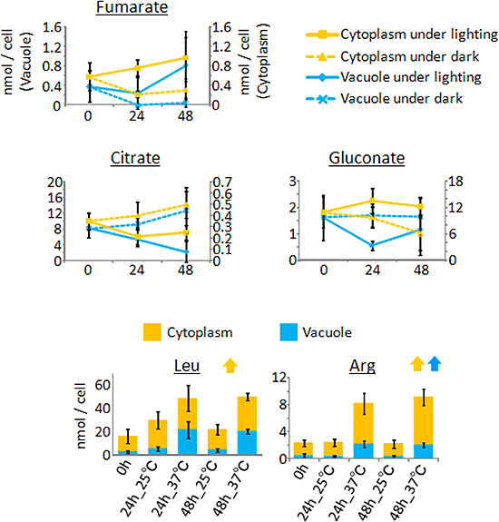
Figure 3: Fluctuation of metabolite concentrations in the cell under varying lighting and temperature conditions.
Top: Concentrations of metabolites under continuous light and dark conditions. Fumarate concentration increased under light conditions (solid line), and decreased under dark conditions (dashed line). Fluctuations of citrate concentration under light conditions were opposite those of fumarate. Gluconate exhibited more complicated concentration fluctuations.
Bottom: Concentrations of metabolites under high (37 °C) and normal (25 °C) temperature conditions. Although the concentrations of both leucine and arginine increased under high temperature conditions, the cytoplasmic concentration of leucine showed little fluctuations. On the other hand, arginine concentration increased in both the vacuole and cytoplasm. These results indicated that fluctuations of metabolite concentrations in a cell were separately regulated for each metabolite.