May 2, 2013 Press Release Biology Physics / Astronomy
New imaging technique to visualize bio-metals and molecules
Metal elements and molecules interact in the body but visualizing them together has always been a challenge. Researchers from the RIKEN Center for Life Science Technologies have developed a new molecular imaging technology that enables them to visualize bio-metals and bio-molecules simultaneously in a live mouse. This new technology will enable researchers to study the complex interactions between metal elements and molecules in living organisms.
Metal elements such as zinc, iron and copper are present in trace amounts in the body and play an important role in myriad biological processes including gene expression, signal transduction and metabolic reactions. Abnormalities in the behaviour of these elements often reflect abnormalities in associated bio-molecules and studying them together can offer great insight into many biological processes.
Bio-molecules can be visualized in living organisms using positron emission tomography (PET), a widely used nuclear medical molecular-imaging technique.
Dr. Shuichi Enomoto, Dr. Shinji Motomura and colleagues, from the RIKEN Center for Life Science Technologies have developed a gamma-ray imaging camera enabling them to detect the gamma-rays emitted by multiple bio-metal elements in the body and study their behavior.
Their second prototype of the system, called GREI–II and presented today in the Journal of Analytical Atomic Spectrometry, enables them to visualize multiple bio-metal elements more than 10 times faster than before, and to do so simultaneously with positron emission tomography (PET).
In the study, the researchers were able to visualise two radioactive agents injected in a tumor-bearing mouse, as well as an anti-tumor antibody labelled with a PET molecular probe agent, simultaneously in the live mouse.
This new revolutionary technology is expected to offer new insights into the relationships between bio-metal elements and associated bio-molecules, and the roles they play in diseases such as diabetes and cancer.
Reference
- Shinji Motomura, Youske Kanayama, Makoto Hiromura, Tomonori Fukuchi, Takahiro Ida, Hiromitsu Haba, Yasuyoshi Watanabe, and Shuichi Enomoto.“Improved imaging performance of semiconductor Compton camera GREI makes for a new methodology to integrate bio-metal analysis and molecular imaging technology in living organisms”. Journal of Analytical Atomic Spectrometry, 2013, doi: 10.1039/C3JA30185K
Contact
Shuichi Enomoto
Next-generation Imaging Team
Imaging Application Group
Division of Bio-function Dynamics Imaging
RIKEN Center for Life Science Technologies
Jens Wilkinson
RIKEN Global Relations and Research Coordination Office
Tel: +81-(0)48-462-1225 / Fax: +81-(0)48-463-3687
Email:pr@riken.jp

Photograph of GREI-II
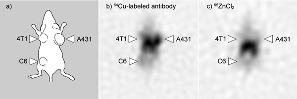
Results of imaging experiment on a tumor-bearing mouse
a) localization of the three lines of tumour cells
b) localization of the labeled antibody
c) localization of Zn-containing injected radioactive agent