May 15, 2013 Press Release Biology
Getting a grip on sleep
All mammals sleep, as do birds and some insects. However, how this basic function is regulated by the brain remains unclear. According to a new study by researchers from the RIKEN Brain Science Institute, a brain region called the lateral habenula plays a central role in the regulation of REM sleep. In an article published today in the Journal of Neuroscience, the team shows that the lateral habenula maintains and regulates REM sleep in rats through regulation of the serotonin system. This study is the first to show a role of the lateral habenula in linking serotonin metabolism and sleep.
The lateral habenula is a region of the brain known to regulate the metabolism of the neurotransmitter serotonin in the brain and to play a key role in cognitive functions.
“Serotonin plays a central role in the pathophysiology of depression, however, it is not clear how abnormalities in regulation of serotonin metabolism in the brain lead to symptoms such as insomnia in depression,” explain Dr. Hidenori Aizawa and Dr. Hitoshi Okamoto who led the study.
Since animals with increased serotonergic activity at the synapse experienced less REM sleep, the researchers hypothesized that the lateral habenula, which regulates serotonergic activity in the brain, must modulate the duration of REM sleep.
They show that removing the lateral habenula in rats results in a reduction of theta rhythm, an oscillatory activity that appears during REM sleep, in the hippocampus, and shortens the rats’ REM sleep periods. However, this inhibitory effect of the lateral habenular lesion on REM sleep disappears when the serotonergic neurons in the midbrain are lesioned.
The team recorded neural activity simultaneously in the lateral habenula and hippocampus in a sleeping rat. They find that the lateral habenular neurons, which fire persistently during non-REM sleep, begin to fire rhythmically in accordance with the theta rhythm in the hippocampus when the animal is in REM sleep.
“Our results indicate that the lateral habenula is essential for maintaining theta rhythms in the hippocampus, which characterize REM sleep in the rat, and that this is done via serotonergic modulation,” concludes Dr Aizawa.
“This study reveals a novel role of the lateral habenula, linking serotonin and REM sleep, which suggests that an hyperactive habenula in patients with depression may cause altered REM sleep,” add the authors.
Reference
- Hidenori Aizawa, Shin Yanagihara, Megumi Kobayashi, Kazue Niisato, Takashi Takekawa, Rie Harukuni, Thomas J. McHugh, Tomoki Fukai, Yoshikazu Isomura, Hitoshi Okamoto. "The synchronous activity of lateral habenular neurons is essential for regulating hippocampal theta oscillation". Journal of Neuroscience, 2013
Contact
Hitoshi Okamoto
Laboratory for Developmental Gene Regulation
RIKEN Brain Science Institute
Jens Wilkinson
RIKEN Global Relations and Research Coordination Office
Tel: +81-(0)48-462-1225 / Fax: +81-(0)48-463-3687
Email: pr@riken.jp
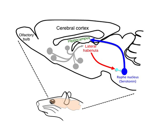
The habenular neural circuit examined in the current study
Lateral view of the rate brain showing how the lateral habenula (red) influences other brain regions such as the hippocampus (green) by way of modulation of the serotonergic activity (blue). Serotonergic neurons connect with many brain regions including the hippocampus. Activation of the lateral habenula inhibits serotonergic neurons in the raphe nucleus (blue) through activation of inhibitory interneurons (light blue).
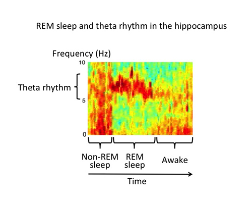
REM sleep and theta rhythm in the hippocampus
Power spectrogram showing changes in the neural activity of the hippocampus during sleep. Frequencies are plotted on the vertical axis with colors ranging from blue (lower) to red (higher). Sleep stages such as awake, REM and non-REM sleep could be identified depending on changes in neural activity. Theta rhythm with 5-8 Hz oscillatory activity is dominant during REM sleep.
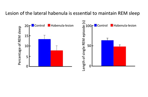
Lesion of the lateral habenula reduces the REM sleep length
Bar graphs showing the percentage of REM sleep (left graph) and the length of single REM episodes in the animals with sham- (control, blue) and lesion-operated rats (Habenula-lesion, red). After sham- and lesion-surgery, hippocampal activity and electromyogram were recorded to determine the sleep stage. Destruction of the lateral habenula significantly reduces these two values suggesting that the lateral habenula is essential for the maintenance of REM sleep.