Aug. 28, 2015 Press Release Biology
Getting a picture of the molecules in a cell in just minutes
Understanding exactly what is taking place inside a single cell is no easy task. For DNA, amplification techniques are available to make the task possible, but for other substances such as proteins and small molecules, scientists generally have to rely on statistics generated from many different cells measured together. Unfortunately, this means they cannot look at what is happening in each individual cell.
Now, thanks to seven years of work done at the RIKEN Quantitative Biology Center and Hiroshima University, scientists can take a peek into a single plant cell and—within minutes—get a view of the small molecules, including metabolites, hormones, nutrients, and lipids, inside it.
In this method, published in Nature Protocols, the contents of the single target cell are directly sucked up by a metal coated glass capillary called a "nanospray tip" under a stereo microscope and the contents are directly fed into the inlet of a mass spectrometer. Within minutes, the mass spectrometer detects hundreds or thousands of molecular peaks, and these peaks can then be matched to databases to determine which metabolites are present in the plant cell under the specific conditions it faced at the time the contents were removed.
This method promises not only to speed up the ability to understand how molecules are distributed in cells in time and space, but also to change how research is done in agricultural science. According to corresponding author Tsutomu Masujima, who leads RIKEN's Single Cell Project, "Using this method we can compare the molecular peaks of cells which are in different stages of growth, different locations, or responding to different circumstances using statistical analyses of the mass spectrometry data. If, for example, we find that certain metabolites are increased in a specific strain, it implies that the enzyme or protein of this specific metabolic pathway may be the key to the specificity of this strain. It might also help us to identify new pathways that are important."
For the moment, the team is focusing on plant cells, but they are preparing a protocol for animal cells—which is much more difficult since the cells are smaller and softer—together with high content screening applications.
Given the potential of the new method, the authors have decided to publish the protocols to the global research community.
Reference
- Takashi Fujii Shuichi Matsuda, Mónica Lorenzo Tejedor, Tsuyoshi Esaki, Iwao Sakane, Hajime Mizuno, Naohiro Tsuyama, and Tsutomu Masujima, "Direct Metabolomics for Plant Single Cells by Live Single Cell Mass Spectrometry", Nature Protocols, doi: 10.1038/nprot.2015.084
Contact
Team Leader
Tsutomu Masujima
Laboratory for Single Cell Mass Spectrometry
Cell Dynamics Research Core
RIKEN Quantitative Biology Center
Jens Wilkinson
RIKEN Global Relations and Research Coordination Office
Tel: +81-(0)48-462-1225 / Fax: +81-(0)48-463-3687
Email: pr@riken.jp
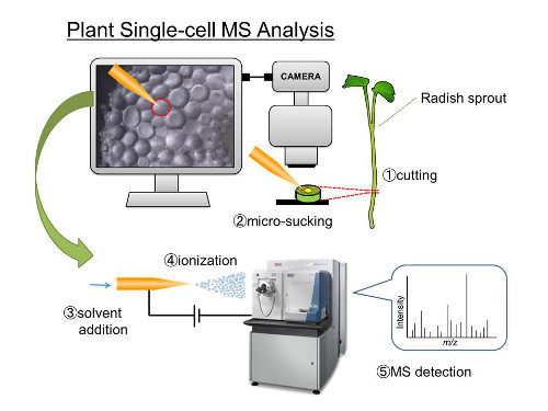
Schematic of the process
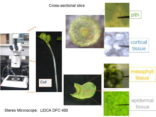
Taking samples from cross-sectional slices