Dec. 25, 2020 Perspectives Biology
Hijacking hibernation
In the last few years, scientists have been able to clarify the energy-saving mechanisms at work in hibernation, culminating in two important papers published in June 2020. But what does this mean for human hibernation?
 Image of a sleeping mouse © MediaProduction / Getty Images
Humans aren’t hibernators. However, we have very recently discovered how to induce hibernation in non-hibernators such as rats and mice, the latter of which normally only go into temporary torpors. Are humans next? Maybe. Another primate, the fat-tailed dwarf lemur native to Madagascar, enters this energy-saving state for up to seven months a year, and at least three other lemur species are known to hibernate. While tucked safely into a tree hollow, this lemur lowers its set body temperature and its metabolic activity plummets by nearly 90%1. Complex physiological adjustments mean that essentials, such as muscle cell activity, are maintained and the lemurs don’t waste away. Studies on the transcriptomes of hibernating bears have revealed complex hibernation-related adjustments occur on the cellular level throughout the year2, so long bouts of human hibernation are a little while off.
But hibernation still holds some exciting possibilities for medical science. For example, hibernating squirrels use melatonin to protect their cells when blood flow increases after months of inactivity. In the early 2000s, a University of Nebraska team used this as inspiration for a treatment to reduce tissue damage when blood suddenly returns after trauma-related hemorrhagic shock. After recent success in rats3 and pigs4, the Nebraska team is now looking at clinical trials. Short bouts of hibernation could also be helpful in limiting tissue damage in stroke victims or organ transplants.
On 11 June 2020, we took a step closer to this when our understanding of hibernation leapt forward. Two studies, both published in Nature5,6, have identified several specific neuron subsets behind the end of the optic nerve that, when activated, lower set body temperatures in mice, inducing an energy-saving, hibernation-like state.
These studies, one of which I co-led, both support modifications to the previous model of thermoregulation activation in the brain. This change to the model has been brewing since 2016 due to a small flurry of studies on the genetic markers involved in the body’s response to skin temperature changes. The studies revealed that the key neurons that cause the sudden drop in body temperature seen in hypothermia are not located in the hypothalamus’ broader medial preoptic area, as previously thought, but in the much smaller median preoptic nucleus7,8,9. This greatly refined our understanding of the neurons and neuropeptides that might be involved in thermoregulation, which is the central mechanism of hibernation.
Burdened by body heat
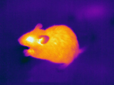 The researchers observed that QFRP-neuron-stimulated mice were comfortable
below 30 degrees Celsius (86 degrees Fahrenheit), when normally they would be cold. This suggested that their set body temperature had been lowered in a manner reminiscent of a deep hibernation state. © EDWARD KINSMAN / SCIENCE PHOTO LIBRARY
The researchers observed that QFRP-neuron-stimulated mice were comfortable
below 30 degrees Celsius (86 degrees Fahrenheit), when normally they would be cold. This suggested that their set body temperature had been lowered in a manner reminiscent of a deep hibernation state. © EDWARD KINSMAN / SCIENCE PHOTO LIBRARY
Thermoregulation is key to energy consumption. Most mammals have a narrow body-temperature range around a set point of 37 degrees Celsius (98.6 degrees Fahrenheit). That’s why even a one-degree rise in humans is our first clue to a fever. In shorter daily torpors, some animals can lower their body temperature closer to ambient air temperature, which is thought to achieve as much as a 70% reduction in energy use. However, in this daily torpor, the animal’s set-point temperature remains the same, and the animals may be enduring a feeling of coldness for the energy-saving benefits10. Deeper hibernation states, most often seen in animals living in extreme environments, reduce an animal’s set-point body temperature for longer, lowering energy use by 90% or more, but this requires much more sophisticated physiological adjustments.
Mice can go into short energy-conserving daily torpors in response to a lack of food. But the aspect of our June study that caused excitement was that by activating the small subset of neurons that express pyroglutamylated RF-amide peptide (QRFP) in mice, we were able to achieve a longer dormancy and a lowered set-point body-temperature, reminiscent of hibernation states5. The other study I mentioned was led by researchers at the Harvard Medical School in the United States, and it echoed our findings, observing shorter torpor states in mice after drug-induced activation of these same neurons6. While we genetically modified our mice to achieve the artificial activation and subsequent hibernation state, the hope is that we have found something that could eventually result in the development of hibernation state control in mammals, and ultimately humans.
Medical synthetic hibernation
The origins of our successful activation of hibernation-like state in mice lie in the long-time study of neuropeptides by wakefulness expert and the corresponding author on our paper, Takeshi Sakurai, and his group at the University of Tsukuba. Sakurai has long been examining functions in the brain of G-protein-coupled receptors, a diverse group of message carriers that are the targets of up to half of all marketed drugs. He observed that injecting QFRP into the brains of mice—QFRP has been linked to feeding and adrenal responses—made them very active. However, when his group tried exciting the neurons that express QRFP in 2017, their mice became still and their body temperature dropped.
Sakurai called me soon after, and my Kobe lab soon began recording the metabolism of the genetically modified mice with a special G protein-coupled receptor expressed on QRFP-neurons, which helped us to stimulate them. With these mice, we were able to induce more than 48 hours of a hibernation-like state within 30 minutes of injecting into the stomach compounds with ligands to the G protein-coupled receptor, a speed that bodes well for the possibilities of use in medical triage. For 48 hours or more, we observed the body temperature of our QFRP-neuron-stimulated mice lowered below 30 degrees Celsius (86 degrees Fahrenheit) and it even dropped to 24 degrees Celsius (75 degrees Fahrenheit) under cold conditions. Their heart rate, oxygen consumption and respiration rate were also all reduced. And we have also been able to replicate this in rats, a species that neither hibernates nor has daily torpor.
In the future, slowing metabolic processes like this might give stroke, heart attack and trauma victims more time before tissue damage becomes too severe to treat. Dormancy could also extend the viability of organ transplants. Currently, a waiting kidney or liver can be preserved in cold solutions for only 24 hours, while a heart or a lung is viable for a mere four to six hours. If we could slow the degradation rate by 90% that would be huge. With an eye to this, the next step for us at RIKEN is to try to induce hibernation in human iPS cell-derived tissues. If we can do this without modifying the genes involved, the possibilities of using short-term dormancy for medicine becomes more real.
Long human hibernation
In terms of longer hibernation states, things become more complex. The fat tissue of grizzly bears has been shown to be among the most affected by hibernation. A 2019 transcriptional analysis showed 900 differentially expressed genes in a hibernating grizzly’s adipose tissue compared to an active season. Hibernation was also characterized by reduced expression of genes associated with insulin signaling, muscle protein degradation, and urea production, and increased expression within muscle protein anabolic pathways. It’s thought that we contain the genes that make this possible, but activating these supporting processes in humans would likely be hugely complex.
However, grizzly bears also become resistant to insulin during hibernation, but they don’t suffer from fluctuating blood sugar levels. Huge weight loss occurs as par for the course, but no bone loss or atrophied muscles have been observed. A human experiencing some of these conditions could easily end up with diabetes, obesity or worse. Will mimicking grizzly bear fat become a future treatment for diabetes? Is aging slowed by hibernation? Could we enter torpor for a few hours every night as part of a beauty or health regime? One theory suggests that deep meditation is designed to replicate hibernation. Can we achieve a hibernation or torpor state using only the mind? These are some of many questions that will be fascinating to explore now that we can induce hibernation states in one of our main mammalian research models.
Eventually, it’s possible that long-term hibernation in humans could be a game changer for space travel, healthcare and even climate change. And like genomic medicine, which once seemed so far away, there will be ethical considerations. What does consent look like to a hibernating human? Will hibernators pay taxes? And when are you really terminally ill? We need to start exploring these questions, as the last year has shown us that the science in this area can rapidly jump forward. If long-term hibernation is possible, it could change our concept of time entirely: could we hibernate through a pandemic, or hit pause and wait for the planet to repair itself? These are questions that our children may have to answer, which is an exciting thought.
References
- 1. Blanco M. B., Dausmann K. H., Faherty S. L. & Yoder A. D. Tropical heterothermy is “cool”: The expression of daily torpor and hibernation in primates. Evol. Anthropol. 27, 147–161(2018). doi: 10.1002/evan.21588
- 2. Jansen, H. T., Trojahn, S., Saxton, M. W. et al. Hibernation induces widespread transcriptional remodeling in metabolic tissues of the grizzly bear. Commun. Biol. 2, 336 (2019). doi: 10.1038/s42003-019-0574-4
- 3. Perez de Lara Rodriguez, C. E., Drewes, L. R. & Andrews, M. T. Hibernation-based blood loss therapy increases survivability of lethal hemorrhagic shock in rats. J. Comp. Physiol. B 187, 769–778 (2017). doi: 10.1007/s00360-017-1076-7
- 4. Wolf, A., Mulier, K. E., Muratore, S. L. et al. D-β-hydroxybutyrate and melatonin for treatment of porcine hemorrhagic shock and injury: a melatonin dose-ranging study. BMC Res. Notes 10, BMC Res. Notes. doi: 10.1186/s13104-017-2975-0
- 5. Takahashi, T. M., Sunagawa, G. A., Soya, S. et al. A discrete neuronal circuit induces a hibernation-like state in rodents. Nature 583, 109–114 (2020). doi: 10.1038/s41586-020-2163-6
- 6. Hrvatin, S., Sun, S., Wilcox, O. F. et al. Neurons that regulate mouse torpor. Nature 583, 115–121 (2020). doi: 10.1038/s41586-020-2387-5
- 7.Machado, N. L. S., Bandaru, S. S., Abbott, S. B. G. & Saper, C. B. J. EP3R-expressing glutamatergic preoptic neurons mediate inflammatory fever. Neurosci 40, 2573–2588 (2020). doi: 10.1523/jneurosci.2887-19.2020
- 8. Yu, S., Qualls-Creekmore, E., Rezai-Zadeh, K., Jiang, Y., Berthoud, H. R. et al Glutamatergic preoptic area neurons that express leptin receptors drive temperature-dependent body weight homeostasis. J. Neurosci. 36, 5034–5046 (2016). doi: 10.1523/jneurosci.0213-16.2016
- 9.Wang, T. A., Teo, C. F., Åkerblom, M., Chen, C., Tynan-La Fontaine, M. et al Thermoregulation via temperature-dependent PGD2 production in mouse preoptic area. Neuron 103, 309–322.e7 (2019). doi: 10.1016/j.neuron.2019.04.035
About the Researchers
Genshiro Sunagawa, Senior Scientist, Laboratory for Molecular Biology of Aging and Laboratory for Retinal Regeneration, RIKEN Center for Biosystems Dynamics Research

Genshiro Sunagawa has been working in the Laboratory for Retinal Regeneration since 2015, where he focuses on active hypometabolism in mammals. He has been working as a clinician (pediatrician and anesthetist) since 2001. The discovery of hibernating tropical primates by Kathrin Dausmann in 2004, inspired him to take a step in the field of biomedical applications of natural phenomena such as hibernation. He hopes to develop safe human hibernation for use in clinical settings.