Jan. 19, 2024 Research Highlight Biology Chemistry
Hot on the tracks of extracellular vesicles
A new technique provides a more comprehensive picture of how the proteins involved in cellular processes communicate via extracellular vesicles

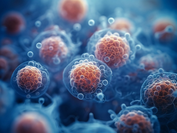
Figure 1: Human cells secreting tiny extracellular vesicles. A novel method developed by RIKEN researchers can identify the proteins involved in the uptake of extracellular vesicles by recipient cells. © JUAN GAERTNER/SCIENCE PHOTO LIBRARY
A new method offers a global view of the proteins responsible for cellular uptake of small extracellular vesicles (EVs)1. As well as offering insights into how healthy cells communicate, it could also reveal how cancer cells spread.
One way that cells communicate with each other is through the secretion and uptake of extracellular vesicles. EVs convey a multitude of cargoes, including proteins, lipids and nucleic acids. Their uptake affects the function of recipient cells by influencing signaling processes and gene expression.
However, despite extensive study of EVs, little is known about their cell-specific uptake by recipient cells.
“Understanding how recipient cells take up EVs is critical for deciphering the broader mechanisms governing cell-to-cell communication in cellular processes in both health and disease,” explains Koshi Imami of the RIKEN Center for Integrative Medical Sciences.
Imami and colleagues have now developed a novel method to track the interaction between EVs and recipient cells. The TurboID-EV system works by labeling recipient cellular proteins close to EVs with biotin (vitamin B7).
“Unlike traditional techniques that tag EVs with fluorescent proteins or use microscopy, our method provides a global view of proteins involved in EV uptake and interactions within recipient cells,” says Imami.
By identifying the biotin-tagged proteins using biochemical enrichment and mass spectrometry, researchers can glean clues into the molecular mechanisms underlying EV uptake.
Imami and colleagues expressed a biotin ligase that is engineered to fuse with EV membranes in human embryonic kidney cells without interfering with EV secretion. By collecting secreted TurboID-EVs and incubating them with recipient cells labeled with heavy amino acids and supplemented with biotin, they could examine biotinylation events that occur during the uptake of EVs.
The researchers identified more than 450 biotinylated recipient proteins. They included well-known ones involved in the process by which cells engulf external substances to bring them in. The team also found proteins involved in intracellular transport and membrane-associated proteins, which could be key for EV uptake in this model.
The method can be adapted for different EV subtypes and cell types. “The versatility of our system allows researchers to investigate the specificity of EV uptake mechanisms in many biological contexts,” Imami says.
Discovering the proteins involved in EV uptake could further our understanding of how cancer cells spread and help develop EV-based drug-delivery systems that target specific cell types.
Imami’s team is now trying to apply the TurboID-EV system to a mouse model to understand how cancer spreads between organs. “Tumor-derived EVs are known to be taken up by organ-specific cells to prepare for cancer to spread to new organs,” Imami explains. “We want to characterize the function of these EVs.”
Related contents
- Halting liver cancer with a sugar look-a-like
- Sugar chain complexes affect cancer cell binding ways
- Protein transport by ‘hug-and-kiss’
Rate this article
Reference
- 1. Li, Y., Kanao, E., Yamano, T., Ishihama, Y. & Imami, K. TurboID-EV: Proteomic mapping of recipient cellular proteins proximal to small extracellular vesicles. Analytical Chemistry 95, 14159–14164 (2023). doi: 10.1021/acs.analchem.3c01015
