Jan. 20, 2012 Research Highlight Biology
Channeling into cell control
A new model of intracellular signaling via calcium ions will assist in understanding the effects of calcium fluctuations
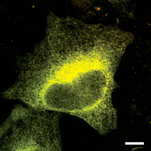 Figure 1: A cell emitting fluorescent signals as a result of attaching specialized proteins to two of its channel-forming IP3Rs (scale bar, 10 µm). Reproduced from Ref. 1 © 2011 the National Academy of Sciences USA
A research team from the RIKEN Brain Science Institute in Wako has visualized and accurately modeled the molecular changes that open and close the internal membrane channels for calcium ions within cells1. The ions moving through these channels act as intracellular messengers, relaying information that regulates the activity of the proteins that control many critical processes of life and death—from fertilization through to development, metabolism and, ultimately, death.
Previous work by the team showed that inositol trisphosphate (IP3) and calcium ions are involved in regulating channel opening and closing. The channels are formed from complexes of four IP3 receptors (IP3R) that bind IP3 and calcium. At low concentrations of calcium ions, channel opening is stimulated; but at higher levels, it is inhibited. Although cell biologists have proposed models depicting this process, they had failed to collect any definitive evidence supporting a particular the mechanism, until now.
In live cells, Takayuki Michikawa, Katsuhiko Mikoshiba and their colleagues attached fluorescent proteins to two of the channel-forming IP3Rs because these receptors change shape in response to the binding of IP3 and calcium, and energy flows between this pair of proteins in a process known as Förster resonance energy transfer (FRET) (Fig. 1). In a detectable way, FRET changes the fluorescent light emitted, so the impact of such links on the conformation of the channel can be studied.
The researchers found there were at least five binding sites on each IP3R, one for IP3 and at least four for calcium. Binding IP3 tended to bring the receptors forming the channel closer together, while calcium tended to make them relax. But the effects of combining the two were not simply additive. At a constant level of IP3, they observed an optimum concentration of calcium that had the most impact on opening the channel.
From these results, the researchers proposed a model whereby IP3 and calcium ions compete with one another—the binding of IP3 prevents calcium linking to certain sites, and vice versa. High concentrations of calcium prevent IP3 from binding at all. Further, the researchers proposed two different types of calcium binding sites: low-affinity sites responsible for channel activation, and high-affinity sites for inactivation.
“During the past five years, we have succeeded in visualizing IP3 dynamics and calcium pump activity,” Michikawa and Mikoshiba say. “In combination with the model for the calcium release channel described in this study, we are now ready to understand what happens in living cells during calcium ion oscillations.”
References
- 1. Shinohara, T., Michikawa, T., Enomoto, M., Goto, J.-I., Iwai, M., Matsu-ura, T., Yamazaki, H., Miyamoto, A., Suzuki, A. & Mikoshiba, K. Mechanistic basis of bell-shaped dependence of inositol 1,4,5-trisphosphate receptor gating on cytosolic calcium. Proceedings of the National Academy of Sciences USA 108, 15486–15491 (2011). doi: 10.1073/pnas.1101677108