Dec. 7, 2012 Research Highlight Medicine / Disease
Searching for the social brain
Imaging of marmoset brains provides an insight into the brain regions involved in regulating social behavior
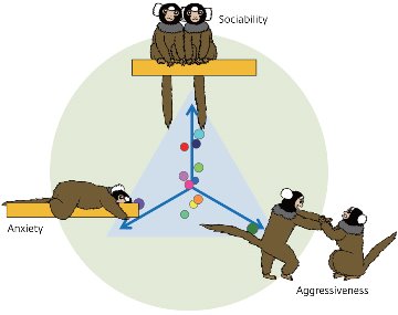 Figure 1: Behavioral traits of common marmosets were identified by their actions during encounters with an unfamiliar animal. Circles represent individual marmosets characterized with three traits. © 2012 C. Takeda, C. Yokoyama and H. Onoe, RIKEN Center for Molecular Imaging Science
Humans exhibit complex social behavior. Understanding the brain function underlying this behavior would help to explain how it goes wrong in social disorders. Evidence shows that the neurotransmitter serotonin is involved in regulating emotional and social behaviors in humans and other primates, particularly negative traits such as anxiety and aggression. It has also been implicated in the formation of social behavioral traits such as social affiliation, dominance and openness.
Despite this, no direct link has been made between such behaviors and the action of serotonin in specific brain regions. Most work has focused on the primitive regions of the brain, such as the amygdala, which is involved in negative emotions. But there is also some evidence for involvement of the cerebral cortex, the outer layer of the brain responsible for higher cognitive function.
A team led by Hirotaka Onoe at the RIKEN Center for Molecular Imaging Science, Japan has studied the brain and behaviors of common marmosets to show that specific regions of the midline cortex, close to the center-line of the brain, are indeed crucial in the formation and regulation of social behavior traits1. Onoe says it is the first study of non-invasive brain functional imaging in conscious marmosets that has been analyzed in combination with measurements of the animals’ social behavior.
The social brain
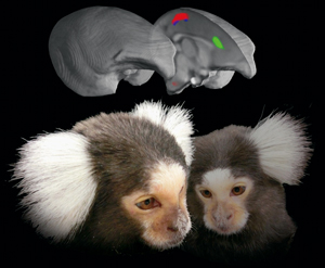 Figure 2: Specific brain regions represent the link of the social traits with serotonin transporter binding revealed by PET imaging. Aggressiveness (red), sociability (blue) and anxiety (green) traits are localized in the medial part of the cerebral cortex. © 2012 C. Yokoyama and H. Onoe, RIKEN Center for Molecular Imaging Science
The researchers started by observing and classifying behavioral traits displayed by the male marmosets when they encountered an unfamiliar male. They identified three traits: aggressiveness, anxiety and sociability (Fig. 1). The combination of these traits expressed in an individual can be considered equivalent to ‘personality’ in humans. The team then moved on to see how serotonin function in the brain related to these traits.
To do this, they measured levels of the serotonin transporter protein (SERT) in the brains of the same marmosets. This protein transports serotonin back into cells after it has been released from neurons. High levels of SERT correspond to high levels of serotonergic nerve terminals in the same region. Measurements were made by injecting a radioactive compound that binds to SERT into the marmosets and detecting the radioactivity in their brains by positron emission tomography (PET). Higher levels of SERT in specific regions were revealed by higher levels of radioactivity.
The researchers found that SERT levels in specific regions correlated with the social traits seen in the marmosets (Fig. 2). Low levels of SERT correlated with lower unfriendliness and higher aggression in the posterior cingulate cortex (PCC). In the anterior cingulate cortex (ACC) and the lateral prefrontal cortex (LPFC), low levels of SERT correlated with higher anxiety. These results indicate that the cognitive basis of these traits is localized to these specific subregions of the midline cortex.
The researchers also measured activity in these regions in different social situations. To do this, they again used PET, but this time they used a radioactive compound to measure the level of metabolism across the brain, corresponding to the amount of neural activity. They found that activity in the PCC, but not the ACC, varied according to whether a marmoset remained alone or was put with an unfamiliar male. Using the same approach, they also showed that functional connections of the PCC and ACC with other regions of the brain were altered by different social situations. The differences in activity show that these regions are central to the cognitive basis of social traits.
A model of human personality
The insights add to the evidence that areas of the midline cortex are involved in the regulation of social behavior by serotonin. What’s more, the differences seen between specific areas demonstrate that different aspects of social behavior are localized to these subregions. Common marmosets are known to have complex social behaviors similar to humans, such as a high level of social learning. By conducting these studies in marmosets, the results are therefore directly relevant to our understanding of human social cognition.
“Our approach uses a reliable model of social traits underlying ‘personality’ in humans,” says the lead author, Chihiro Yokoyama. “Our findings confirm the previous knowledge of the relationship between serotonin and social behaviors and further highlight the role of the medial part of the cerebral cortex as the site of action.” The study could lead to a better understanding of complex social disabilities such as depression, anxiety disorders and autism. Using the same approach, Onoe’s team now want to gain a deeper understanding of the action of serotonin in specific brain regions.
“In the next step of this research we would like to elucidate a pivotal role of the serotonergic system in the medial part of the cortex in expressing social behaviors,” Onoe says. “Our method of live imaging provides not only visualization of the neural processes in the living and functioning brain, but can also shed light on the relationship between genes and social behavior.”
References
- 1. Yokoyama, C., Kawasaki, A., Hayashi, T. & Onoe, H. Linkage between the midline cortical serotonergic system and social behavior traits: positron emission tomography studies of common marmosets. Cerebral Cortex advance online publication, 12 July 2012. doi: 10.1093/cercor/bhs196
About the Researcher
Hirotaka Onoe

Hirotaka Onoe was born in Tokyo in 1960. He graduated from the Tokyo University of Pharmacy and Life Sciences in 1983, and in 1993 obtained his PhD in Pharmacology from Osaka University. After working at the Osaka Bioscience Institute as a research scientist for nearly ten years, Onoe held the position of senior researcher at the Tokyo Metropolitan Institute for Neuroscience for nine years from 1998. He joined the RIKEN Molecular Imaging Research Program as Laboratory Head of the Functional Probe Research Laboratory in 2007, and the laboratory was incorporated into the RIKEN Center for Molecular Imaging Science in 2008. His research focuses on the development of molecular imaging probes for positron emission tomography (PET) and neuroscience.