Nov. 6, 2015 Research Highlight Biology
Corralling chromosomes
Chromosomes attach to microtubules later than expected during egg formation, which may increase the chances of erroneous attachments forming
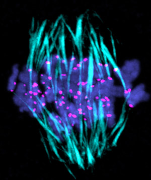
 Figure 1: Unlike during mitosis, in meiosis chromosomes are stretched apart before kinetochore–microtubule attachments are stabilized. This could increase the possibility of erroneous attachments forming. Reprinted from Ref. 1, Copyright 2015, with permission from Elsevier.
By analyzing chromosome dynamics during cell division in live mouse oocytes, RIKEN researchers have discovered that the paired chromosomes attach to filaments called microtubules only after they have started to stretch apart from each other toward opposite poles of the cell1. This later-than-expected microtubule attachment increases the possibility of erroneous attachments forming.
Cells divide and reproduce in two ways, mitosis and meiosis. Mitosis produces two identical daughter cells, whereas meiosis gives rise to four sex cells.
During mitosis, chromosomes replicate and separate from each other, forming two daughter cells that possess the same genetic information as the parent cell. When the chromosomes separate, they attach to microtubules in a specialized chromosomal structural region known as the kinetochore. Destabilizing enzymes called Aurora B and C then eliminate any incorrect kinetochore–microtubule attachments. When all the kinetochores are correctly attached, the chromosomes stretch apart and separate. However, it was unclear to what extent these mitosis processes govern chromosomal behavior during meiosis.
Now, Shuhei Yoshida, Masako Kaido and Tomoya Kitajima at the RIKEN Center for Developmental Biology have used time-lapse microscopy to analyze the sequence of events that chromosomes undergo in mouse oocytes during the first phase of meiosis. In this stage, paired chromosomes that have replicated their genetic information align with each other as well as with microtubules. They then get pulled apart by the microtubules. Importantly, the researchers found that, in contrast with mitosis, during meiosis chromosomes are stretched apart before kinetochore–microtubule attachments are stabilized (Fig. 1).
Correct kinetochore–microtubule attachment is crucial for accurate segregation of chromosomes. “Our finding that stretching occurs before stable attachments are formed suggests that oocyte chromosomes are inherently prone to making microtubule attachment errors,” explains Kitajima.
Kitajima and his colleagues found that Aurora B and C were localized to sites of kinetochore–microtubule attachment in meiotic oocytes even after the chromosomes were stretched apart. While this seems to go against the accepted view that Aurora B and C limit incorrect attachments during meiosis, the team found that inhibiting Aurora B and C caused the number of correct attachments to increase. Therefore, Aurora B and C prevent stabilization of correct attachments after the stretching phase.
In further studies, the team plans to investigate a new question raised by their research. “This study has revealed how chromosomes can stretch without stable microtubules,” says Kitajima. “We now intend to explore the underlying mechanism of this process.”
References
- 1. Yoshida, S., Kaido, M. & Kitajima, T. S. Inherent instability of correct kinetochore-microtubule attachments during meiosis I in oocytes. Developmental Cell 33, 589–602 (2015). doi: 10.1016/j.devcel.2015.04.020
