Sep. 15, 2017 Research Highlight Biology
Measuring morphogenesis of organs
A new statistical-based method provides insight into how developing organs morph into their mature shapes
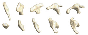 Figure 1: A new statistical method has been used to visualize tissue deformation process during the early development of chick brain tissue. The images shown here are observed morphological changes of apical surfaces of the forebrain (top row: dorsal; bottom row: anterior) to which the proposed method was applied. Reproduced from Ref. 1 and licensed under CC BY 4.0 © 2016 Y. Morishita et al.
By merely measuring the positions of fewer than one in ten cells in a developing organ, scientists can reconstruct how the entire organ acquires its shape thanks to a statistical analysis method developed by an all-RIKEN team1. This method will advance our understanding of organ growth by overcoming the limitations of traditional imaging.
Due to the difficulty of imaging and charting individual cells in large tissue samples over time, scientists know little about the morphological changes that occur when an organ transforms from its initial sheet-like form to a more complex three-dimensional structure.
“Quantifying how developing tissue changes shape over time—a process called deformation—is a key step in elucidating the mechanisms that determine specific organ structure,” says Yoshihiro Morishita of the RIKEN Quantitative Biology Center. “One of the ultimate goals of studying organ morphogenesis is to understand the relationship between tissue deformation and cellular activity that contributes to growth patterns.”
Morishita and his team have now developed a method that involves labeling a low amount (1–10 per cent) of an organ’s cells with fluorescent tags, measuring their positions before and after deformation, and then conducting statistical analysis to infer the transformation undergone by the organ as a whole. This approach can describe the deformation patterns of sheet-like tissue—the starting morphology of complex organs such as the brain and heart—with an error margin of just a few cells wide.
 Yoshihiro Morishita, RIKEN Quantitative Biology Center © 2017 RIKEN
To validate their method, the researchers accurately mapped the early developmental stages of chick brain tissue (Fig. 1). They uncovered evidence that the chick forebrain assumed its more mature morphology via the positional rearrangement of existing cells rather than the stimulus of new cell growth.
“These results are important not only for purely scientific interest, but also for potential medical advances that come with the ability to design and control the genesis of functional organs from stem cells,” states Morishita. “Recent successes in inducing tissue growth from stem cells have been due in part to the ability of the cells to self-organize. By understanding the underlying dynamics, we will be able to influence these processes ourselves.”
Looking forward, Morishita says his group wants to understand how the development of organ morphology differs between different organs. “Both the brain and the heart begin with the deformation of simple tubular structures, but then develop into completely distinct organs. Knowing what makes these processes tick is one of the ultimate goals of developmental biology.”
Related contents
References
- 1. Morishita, Y., Hironaka, K., Lee, S.-W., Jin, T. & Ohtsuka, D. Reconstructing 3D deformation dynamics for curved epithelial sheet morphogenesis from positional data of sparsely-labeled cells. Nature Communications 8, 15 (2017). doi: 10.1038/s41467-017-00023-7