Oct. 28, 2022 Research Highlight Biology
Revealing the structure of the light-harvesting phycobilisome of cyanobacterium
The structure of light-harvesting antennas in a species of cyanobacteria is revealed, yielding insights into energy transfer mechanisms during photosynthesis
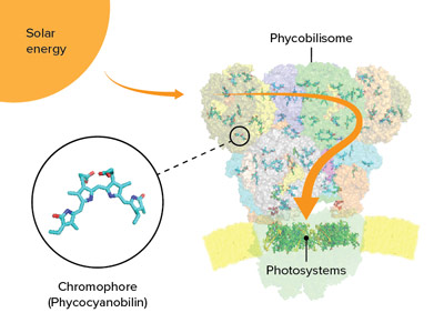 Figure 1: Chromophores interspersed in the phycobilisome of cyanobacteria act as tiny antennas, capturing sunlight. The energy is then conveyed to photosystems at the base of the phycobilisome. © 2022 RIKEN SPring-8 Center
The structure of the ‘antenna’ that a blue-green alga uses to harvest light has been determined by RIKEN researchers and compared with those of four other species1. In addition to providing clues about the evolution and diversity of cyanobacteria, this research could inform the development of efficient photoreactive compounds.
Located on the surface membranes of cyanobacteria (or blue-green algae) and other algae, phycobilisomes are bundles of proteins and chromophores that act as antennas for capturing light during photosynthesis (Fig. 1).
“Phycobilisomes absorb light at wavelengths that are difficult for other light-absorbing photosynthetic proteins, such as photosystems I and II, to utilize,” explains Keisuke Kawakami at the RIKEN SPring-8 Center. “Each algal species has its own unique phycobilisome structure. Gaining insights into these structures will boost our knowledge of efficient energy production from sunlight.”
Phycobilisomes come in five shapes, of which the cyanobacterium called Thermosynechococcus vulcanus has the most common one—a half-sphere with a fan-like arrangement of rods protruding from the core. Light energy is absorbed and then transferred through the rods and cores to photosystems I and II.
Now, by using cryo-electron microscopy, Koji Yonekura, also at the RIKEN SPring-8 Center, and his team have analyzed the structure and function of the phycobilisome of T. vulcanus, and compared it with those of four previously reported species.
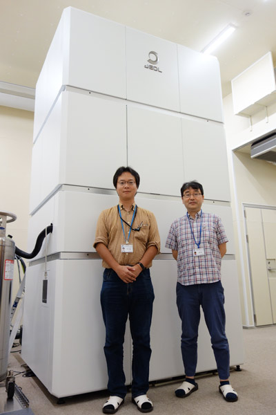 Keisuke Kawakami (left) and Koji Yonekura (right) standing in front of the cryo-electron microscope they used to look at the phycobilisome of the cyanobacterium Thermosynechococcus vulcanus. © 2022 RIKEN
The researchers discovered that the core consists of five cylinders of proteins, rather than the more common three-cylinder structure, and they also identified a new type of cylinder. Each phycobilisome has several rods projecting from the core, formed of stacked proteins and chromophores connected by linker proteins. T. vulcanus only has one type of chromophore, called phycocyanobilin, whereas some algae have more than one.
“Uncovering the structure of linker proteins and amino acids around each phycocyanobilin, and the interactions between them, is critical for understanding unidirectional energy transfer,” says Kawakami. “We were surprised to find that amino-acid residues located around certain phycocyanobilins can manipulate the absorption wavelengths of those chromophores, enabling continuous and rapid energy transfer even under changing light conditions.”
The team also found that these amino-acid residues differ subtly between algal species, probably as a result of the different light conditions in different growth environments.
“Investigating the phycobilisome structures in different species will help us understand the diversity and evolution of algae,” says Kawakami. “Taking inspiration from phycobilisomes could inform the development of optical devices that exploit the mechanism of unidirectional energy transfer using a single chromophore.”
Related contents
- Dual photoreceptor identified in an oceanic green picoplankton
- Structural study of photosystem I containing chlorophyll d provides hints of how it harnesses low-energy light
Rate this article
Reference
- 1. Kawakami, K., Hamaguchi, T., Hirose, Y., Kosumi, D., Miyata, M, Kamiya, N. & Yonekura, K. Core and rod structures of a thermophilic cyanobacterial light-harvesting phycobilisome. Nature Communications 13, 3389 (2022). doi: 10.1038/s41467-022-30962-9