Apr. 14, 2023 Research Highlight Biology
How mouse embryos determine left from right
Resolution of a two-decade debate enhances our understanding of how embryos develop differences between their left and right sides
RIKEN biologists have discovered how tiny hairs in embryos detect flowing fluid, which ultimately leads to the left and right sides of the embryo developing differences1. As well as resolving a long-standing debate, this finding will inform research into disorders that arise when this process malfunctions.
Viewed from the outside, the left and right sides of vertebrate bodies usually appear indistinguishable. But the situation is very different on the inside, with many organs such as the heart, liver and spleen being positioned to either the left or right of the central left–right axis.
By contrast, embryos begin as symmetric bundles of cells. Researchers have long been interested in how differences between the left and right sides develop in embryos.
Tiny hairs, or cilia, in a mouse embryo gyrate clockwise, setting up a leftward flow in the surrounding fluid. This fluid flow is then picked up by static cilia, called immotile cilia, located to the left and right of the moving cilia. This detection of fluid movement causes the left and right sides of the embryo to develop differently.
However, debate surrounds how the immotile cilia sense the fluid flow. Two mechanisms have been proposed: one in which the movement is detected through chemicals in the flow and another in which it is detected via the mechanical force. But no-one had shown experimentally which one is correct until now.
Hiroshi Hamada and Takanobu Katoh of the RIKEN Center for Biosystems Dynamics Research and co-workers have put this debate to rest by showing that the cilia in mice embryos detect the movement mechanically (Fig. 1).
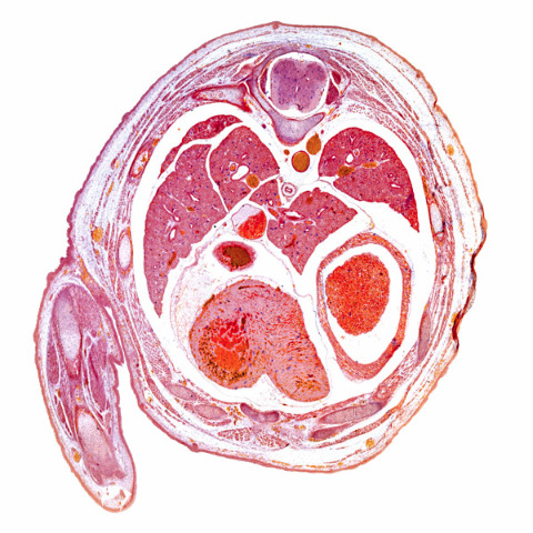
Figure 1: A light micrograph of a section through the thorax of a mouse embryo, which shows left–right asymmetry. RIKEN researchers have discovered how cilia detect fluid flow, which eventually leads to this asymmetry. © DR KEITH WHEELER/SCIENCE PHOTO LIBRARY
Using microbeads trapped in a laser beam, the team manipulated a single cilium and observed a signal involving calcium ions. This showed that mechanical stimulus of just one cilium can determine left–right asymmetry in embryos.
Using advanced microscopy techniques, they also found that left and right cilia are bent in opposite directions. Furthermore, they discovered that the cilia sense the bending direction because channels within them are not symmetrically distributed. Consequently, calcium signals are only triggered when fluid strikes them in one direction, explaining why only cilia on the left side of an embryo are activated.
“Our research has finally answered one of the most important questions for left–right determination, namely why leftward nodal flow activates only the left side,” says Katoh. “This represents an important step towards understanding the mechanism of left–right determination.”
These findings could have practical applications. “Our results provide useful information for studies of how organs form,” says Katoh. “They might also be helpful for finding ways to treat cilia-related disorders.”
Related contents
- mRNA degradation induced by fluid flow breaks left–right symmetry in vertebrates
- Simulations reveal the mechanics of early eye formation in a dish
- Reconstructing the cellular signaling pathways that shape trachea development
Rate this article
Reference
- 1. Katoh, T. A., Omori, T., Mizuno, K., Sai, X., Minegishi, K., Ikawa, Y., Nishimura, H., Itabashi, T., Kajikawa, E., Hiver, S. et al. Immotile cilia mechanically sense the direction of fluid flow for left–right determination. Science 379, 66–71 (2023). doi: 10.1126/science.abq8148