Nov. 13, 2023 Research Highlight Biology
Dissecting the structural secrets of the inactive X chromosome
Sophisticated techniques offer insights into the unusual features of the inactive X chromosome in female mammalian cells
RIKEN cell biologists have provided an unprecedented glimpse into the distinctive features of an unusual chromosome—an inactivated X chromosome copy carried by every female cell1.
Not all X chromosomes are created equal. In every cell of female mammals, one of the two X chromosome copies is compacted into an inactive form known as the inactive X chromosome (Xi). It differs in both structure and function from its active counterpart, and other chromosomes.
Unlike most chromosomes, which replicate gradually throughout the hours-long ‘S phase’ stage of cell division, the Xi is copied in the latter half of this phase. Researchers have hypothesized that this replication behavior is closely tied to the unusual structure of the Xi, which is more uniform and compact than other chromosomes.
“However, this was still uncertain when we started our research,” says Rawin Poonperm of the RIKEN Center for Biosystems Dynamics Research (BDR). “We thought that a detailed analysis of the Xi’s replication timing might uncover its purpose as well as provide new insights into its 3D organization.”
The researchers used two cutting-edge analytical methods to address this question by differentiating cultured embryonic stem cells from mice. The first, which was developed by a group led by Ichiro Hiratani (also of BDR), precisely determined the timing of when specific chromosomal sequences undergo replication. In parallel, they employed a second technique, which revealed the 3D structure of chromatin—the combination of DNA and protein that forms chromosomes—thereby offering insights into gene expression activity.
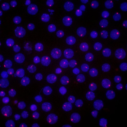
Figure 1: Inactivated X (Xi) chromosomes (red) in cultured cells from mice, which have been labeled with a fluorescent probe for the Xi-specific RNA marker Xist. © 2023 RIKEN Center for Biosystems Dynamics Research
This combination of the two techniques proved highly effective. “Our high-resolution replication analysis revealed that the Xi is copied quickly within the second half of the S-phase as it formed during differentiation. Moreover, our 3D structure results showed that dynamic changes in the replication timing of the Xi closely corresponded to changes in chromatin organization during differentiation,” says Poonperm. She adds that their structural analysis of the Xi also revealed some surprises, including differences in the magnitude of compaction and inactivation at different sites in the chromosome.
When the researchers analyzed differentiated cells that lacked a gene called SmcHD1, which helps keep the Xi inactive, they observed reactivation of normally dormant genes at the edge of the chromosome. Most other Xi genes remained quiescent, however, hinting at hidden structural complexity in this seemingly uniform chromosome.
Hiratani’s group next plans to examine the process of X chromosome inactivation itself. This will entail considerably more challenging experiments in actual mouse embryos rather than cultured embryonic stem cells. But Poonperm sees an exciting opportunity to resolve mysteries that have surrounded this process for decades. “We firmly believe that there are more surprising discoveries to be made,” she says.

Ichiro Hiratani (left), Rawin Poonperm (right) and co-workers have applied sophisticated molecular biology techniques to gain new insights into the unusual features of the inactivated X chromosome copy carried by every female mammalian cell. © 2023 RIKEN
Related contents
- Scientists lay foundation for single-cell level understanding of DNA replication
- How chromosomes change their shape during cell differentiation
Rate this article
Reference
- 1. Poonperm, R., Ichihara, S., Miura, H., Tanigawa, A., Nagao, K., Obuse, C., Sado, T. & Hiratani, I. Replication dynamics identifies the folding principles of the inactive X chromosome. Nature Structural and Molecular Biology 30, 1224–1237 (2023). doi: 10.1038/s41594-023-01052-1