Mar. 13, 2020 Research Highlight Biology
Stem cells exert tight control over the timing of brain development
Insights into mechanisms controlling early brain development could also help scientists understand this organ’s evolution
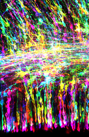
 Figure 1: A fluorescence microscopy image of brain stem cells in the developing ferret brain, which acquires deep folds typically seen in primates and higher mammals. Matsuzaki’s group’s findings on brain stem cell proliferation and development could help explain how such structures form. © 2020 RIKEN Center for Biosystems Dynamics Research
Neural stem cells affect the timing and trajectory of division in brain development more than previously thought, RIKEN researchers have found1. This discovery could have important implications for our understanding of the evolution of the mammalian brain.
“We’ve been interested in the general design principles underlying how brains are built,” says Fumio Matsuzaki of the RIKEN Center for Biosystems Dynamics Research.
Matsuzaki and his team have focused on brain stem cells known as radial glia (Fig. 1). These cells, which give rise to all the neurons in the cerebral cortex, are stretched between two surfaces, known as the apical and basal membranes.
Radial glia initially divide laterally in a symmetric fashion, yielding two daughter radial glia that are also anchored to both surfaces. Over time, some of these cells begin dividing asymmetrically, pushing at the basal surface and causing the developing brain to expand outward. These asymmetric divisions yield more-mature cells that ultimately produce neurons and other brain cells.
For decades, developmental biologists have postulated that this transition is governed by changes in the orientation of the cellular spindles—molecular machines that govern mitotic division—relative to the apical surface.
But previous work by Matsuzaki and his colleagues has called that model into question2,3. “Our findings have provided strong evidence that mitotic spindle orientation does not determine whether self-renewal or differentiation occurs,” he says.
The latest findings by Matsuzaki’s team not only reinforce this hypothesis, but also offer an alternative mechanism to account for this shift.
The researchers manipulated the spindle orientation of radial glia and then imaged what happened. Altered orientation caused radial glia to detach from the apical membrane, but did not necessarily lead to asymmetric division and expansion. Instead, early-stage radial glia have the capacity to extend membrane projections or ‘endfeet’ that can reattach to the apical surface, allowing symmetric division to proceed.
“This ability to regenerate prevents radial glia from detaching and migrating,” explains Matsuzaki. “Only later in development do these detachment events lead to expansion and differentiation.”
This is not just important for brain development; it also has evolutionary consequences since this process governs the expansion of cortical layers near the brain surface and complex folds seen in the brains of primates and other higher mammals.
Matsuzaki is now keen to understand the forces that control when and why radial glia ‘let go’ and begin to differentiate. “We don’t yet know the exact mechanisms by which one daughter cell self-renews while the other differentiates,” he says. “This is an enigma in our field.”
Related contents
- Splitting up cell fates during brain development
- Neurons branch out the right way
- Growing brains in the lab
References
- 1. Fujita, I., Shitamukai, A., Kusumoto, F., Mase, S., Suetsugu, T., Omori, A., Kato, K., Abe, T., Shioi, G., Konno, D. & Matsuzaki, F. Endfoot regeneration restricts radial glial state and prevents translocation into the outer subventricular zone in early mammalian brain development. Nature Cell Biology 22, 26–37 (2020). doi: 10.1038/s41556-019-0436-9
- 2. Shitamukai, A., Konno, D. & Matsuzaki, F. Oblique radial glial divisions in the developing mouse neocortex induce self-renewing progenitors outside the germinal zone that resemble primate outer subventricular zone progenitors. The Journal of Neuroscience 31, 3683–3695 (2011). doi: 10.1523/JNEUROSCI.4773-10.2011
- 3. Konno, D., Shioi, G., Shitamukai, A., Mori, A., Kiyonari, H., Miyata, T. & Matsuzaki, F. Neuroepithelial progenitors undergo LGN-dependent planar divisions to maintain self-renewability during mammalian neurogenesis. Nature Cell Biology 10, 93–101 (2008). doi: 10.1038/ncb1673
