Dec. 28, 2017 Research Highlight Chemistry
Probing water molecules at cell membrane interfaces
Ultrafast laser pulses offer insights into the role water plays in transport through biological membranes
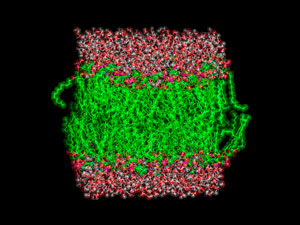 Figure 1: Phosphatidylcholine lipids in biological membranes consist of two parts—a hydrophobic hydrocarbon chain (green) and a hydrophilic phosphate end (pink and red). © LAGUNA DESIGN/ SCIENCE PHOTO LIBRARY
A spectroscopic technique that picks out the signal just from water molecules in thin layers above and below the cell membrane has given RIKEN researchers new insights into the role this water plays in cell processes1.
The thin layers of water on the inner and outer surfaces of cell membranes are vital to managing the flow of ions and small molecules into and out of cells. But it has been hard to study their role because conventional characterization methods cannot distinguish between water at the membrane surfaces and bulk water inside cells.
To overcome this problem, Tahei Tahara at RIKEN’s Molecular Spectroscopy Laboratory and colleagues used a recently developed variation of sum frequency generation spectroscopy—a technique widely used to probe surface layers. It uses ultrashort laser pulses that cause only surface water molecules to vibrate at a cell membrane interface. Consequently, unlike in conventional vibrational spectroscopy, signals from the stretching of oxygen–hydrogen bonds in these few water molecules are not drowned out by signals from the many bulk water molecules. This method enables the up–down orientation of water molecules at the interface to be determined.
The team focused on phosphatidylcholine lipids, a major component of biological membranes. They are known as zwitterions because their heads have both negative (phosphate) and positive (choline) parts (Fig. 1). “Hydration at zwitterionic lipids is directly related to the elementary processes of life at the molecular level,” notes Tahara.
 Tahei Tahara and his team have used an ultrafast laser to probe the role water plays in transporting ions and small molecules through biological membranes. © 2017 RIKEN
To observe the evolution of the hydration process, the team collected spectra every hundred femtoseconds (one femtosecond = 10−15 second). They confirmed that in model biological membranes both the lipid’s phosphate and choline sites have water molecules grouped around them, as the researchers proposed in 2012.
The team also found that water dynamics at the interface are not just the sum of the water molecules surrounding these two parts of the zwitterions. “Water molecules in the two hydration shells begin communicating and all the water molecules in the area start flapping,” Tahara says.
This is surprising since the team had expected the water molecules surrounding each charged site to operate independently of each other. “The motions of the water molecules in two different hydration shells are quite dependent on each other because of the hydrogen bonds between all the water molecules in the vicinity,” explains Tahara.
“This study is just the beginning,” he says. “We still know very little about the dynamics of interfacial molecules.” A deeper understanding of how water behaves on cell membrane surfaces will eventually improve knowledge of biological reactions occurring at cell membranes, he adds.
Related contents
- The hunt for partially hydrated electrons
- Water’s triple play at membrane interfaces
- Imaging lipid rafts reveals some surprises
References
- 1. Inoue, K., Singh, P. C., Nihonyanagi, S., Yamaguchi, S. & Tahara, T. Cooperative hydrogen-bond dynamics at a zwitterionic lipid/water interface revealed by 2D HD-VSFG spectroscopy. The Journal of Physical Chemistry Letters 8, 5160–5165 (2017). doi: 10.1021/acs.jpclett.7b02057