Oct. 10, 2023 Research Highlight Biology
Mapping connections of the marmoset prefrontal cortex
The prefrontal cortex in the marmoset has two distinct types of connections to other cortical areas
A comprehensive map of the marmoset brain by RIKEN neuroscientists has revealed that two types of connections link the prefrontal cortex to other cortical areas1. This finding could provide insight for addressing neurological disorders involving the prefrontal cortex.
A lot of what we know about the human brain comes from experiments on rats and mice. But unlike humans, rodents lack a well-developed prefrontal cortex—the brain region housed immediately behind the forehead that regulates our thoughts, emotions and actions. Knowledge of the prefrontal cortex gained from rodent studies may thus be providing only a partial picture of the human prefrontal cortex.
“One of the most remarkable features of the human brain is its large prefrontal cortex, which is much more evolved and highly specialized than the rodent counterpart,” says Akiya Watakabe of the RIKEN Center for Brain Science (CBS). “It controls the emotions and thoughts, so in a sense it’s a center of higher brain functions.”
Primates such as marmosets provide a helpful stepping stone between rodents and us since their prefrontal cortex is much more developed than rodents’. One thing that was largely unknown is how the prefrontal cortex connects to and interacts with the other brain areas.
Now, Watakabe and Henrik Skibbe, also of the CBS, and co-workers have identified two types of projections—patchy and diffuse ones—that connect the prefrontal cortex to the cortex and striatum. The patchy connections had multiple column-like structures that were smaller than a millimeter in size. In contrast, the diffuse connections spread out widely across the cortex and striatum.
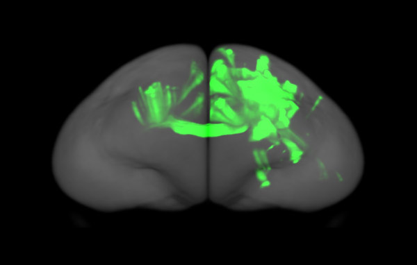
Figure 1: One example of a 3D reconstruction of a tracer data, showing how the prefrontal projections converge to many columns of cortical protrusions in the ipsilateral and contralateral hemispheres of the marmoset brain. © 2023 RIKEN Center for Brain Science
“We uncovered two kinds of connectivities: the patchy one connects two points very strongly with each other, whereas the diffuse one connects to very large regions, but only very loosely,” explains Watakabe. “So we expect that the two connections have different functions.”
The team uncovered this structure by injecting viral tracer into more than 40 locations and then observing the paths they took. It took about seven years to collect and analyze all the data.
This is the first time such a comprehensive and detailed projection map has been generated for primates. “Whole-brain analysis at this high resolution hasn’t been done before for primates because it generates too much data,” says Watakabe. Skibbe took the data generated from the different brains and mapped it onto a common brain template. He also used deep learning to accurately detect the tracer signal for quantitation. “When I saw his processed data, it was so beautiful to look at,” recalls Watakabe.
The whole-brain map of the prefrontal cortex projections in the marmoset is freely available on the Brain/MINDS Data Portal.
Related contents
- Japan’s big brain project: advances light up marmoset brains
- Optical stimulation causes marmosets to move their forelimbs
- Deepening our understanding of selfish behavior
Rate this article
Reference
- 1. Watakabe, A., Skibbe, H., Nakae, K., Abe, H., Ichinohe, N., Rachmadi, M. F., Wang, J., Takaji, M., Mizukami, H., Woodward, A. et al. Local and long-distance organization of prefrontal cortex circuits in the marmoset brain. Neuron 111, 2258–2273 (2023). doi: 10.1016/j.neuron.2023.04.028